
A breast cancer diagnosis does not, on its own, tell us how the disease will progress. Some tumours depend on oestrogen to grow, others produce large amounts of the HER2 protein, and others have neither of these markers. Some respond well to the first treatment; others find ways to escape it.
And there is even more diversity when we look inside the same tumour. Cells are not all alike, nor do they remain unchanged over time. They can change their state, interact with the cells around them or acquire characteristics that help them survive and spread.
This diversity is at the heart of much of the breast cancer research carried out at the centres of the Barcelona Biomedical Research Park (PRBB). The questions range from what happens inside the nucleus of a tumour cell to how we can improve screening, prevent metastasis or better support the health of people who have experienced the disease.
Key points
- Breast cancer includes biologically different types of tumours. Hormone receptors and HER2 help classify them and guide treatment, but they account for only part of their diversity.
- PRBB centres study different stages of the disease: risk and detection, tumour growth, metastasis, treatment resistance and effects that may persist years later.
- A finding in cells or mice, an association observed in a population and the result of a clinical trial do not tell us the same thing. Understanding the stage of research each study represents is essential for interpreting its findings.
One name, different tumours
Among the first things analysed in a breast tumour are its oestrogen and progesterone receptors and the HER2 protein. These markers provide clues about what drives tumour growth and help clinicians select treatments targeting some of these pathways.
When a tumour has neither hormone receptors nor high levels of HER2, it is classified as triple-negative. But even these categories do not explain all the diversity between tumours.
Another part of the answer lies in how cells use their DNA. To activate some genes while keeping others switched off, they must open and close regions of chromatin, the structure formed by DNA and the proteins that organise it.
In 2016, the Chromatin and Gene Expression group led by Miguel Beato at the Centre for Genomic Regulation (CRG) described an unexpected mechanism in hormone-sensitive breast cancer cells in a study published in Science. With Roni Wright as first author, the team discovered that these cells could generate ATP within the nucleus itself, providing the energy needed to reorganise chromatin after receiving a hormonal signal and activate the corresponding genes.
The relationship between metabolism and gene regulation remains an active area of research at the CRG. Sara Sdelci’s group studies enzymes best known for their metabolic roles but which are also found in chromatin. In a study published in 2024 in Nature Communications, the group focused on IMPDH2, a protein that accumulated in the chromatin of triple-negative breast cancer cells and was involved in the response to DNA damage. Altering its activity could lead to the death of tumour cells.
The clinical classification of a tumour is essential, but cells within the same category may use genes, energy and repair pathways in different ways.
Before diagnosis: risk and detection
Not all breast cancer research begins with a tumour. Some studies look much further back, asking which factors are associated with the risk of developing the disease.
In 2018, Cristina O’Callaghan-Gordo, Mark Nieuwenhuijsen and other researchers from the Barcelona Institute for Global Health (ISGlobal) analysed the relationship between green spaces and breast cancer as part of the MCC-Spain study, published that year in International Journal of Hygiene and Environmental Health.
Living closer to urban green spaces was associated with a lower risk of breast cancer, while proximity to agricultural areas showed an association in the opposite direction. This does not mean that living next to a park directly protects against cancer. The findings raise further questions about the differences between these environments and the roles of pollution, physical activity and other exposures.
When it comes to screening, the questions focus on whether we can better identify who is at greater risk between one mammogram and the next.
Marta Román, from the Epidemiology and Evaluation group at the Hospital del Mar Research Institute (HMRIB), contributed to a study combining clinical information with data extracted from mammograms using artificial intelligence. The study was published online in 2025 in European Radiology and used mammograms from 2.193 women participating in the Hospital del Mar screening programme to estimate their risk of being diagnosed with breast cancer over the following two years.
The model combining the different sources of information performed best, although the improvement over some of the other models was small. Before introducing such a tool into a screening programme, it is necessary to confirm that it also works in other populations and genuinely helps improve decision-making.
A tumour does not grow alone
We now know that an important part of the story of cancer unfolds around tumour cells.
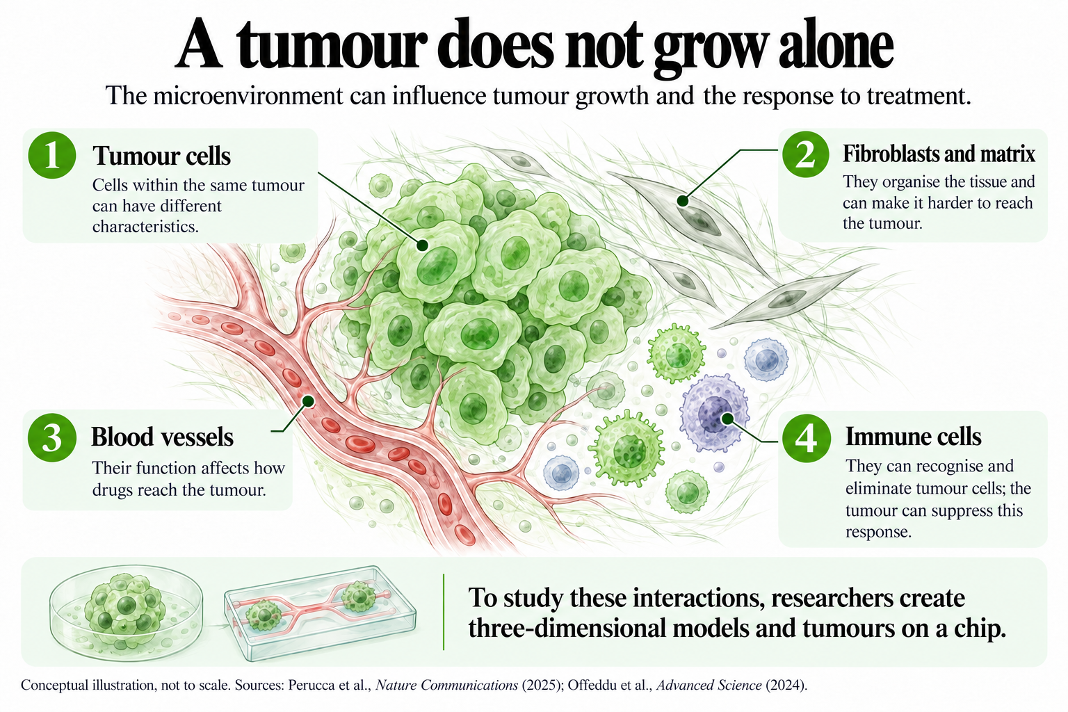
Fibroblasts, immune cells, blood vessels and molecules that form the structure of the tissue coexist with the tumour and make up its microenvironment. This environment can support tumour growth, prevent immune defences from reaching cancer cells or make it harder for a drug to penetrate the tissue.
To observe these interactions under conditions more closely resembling those of a real tumour, Alexandre Calon’s group at HMRIB worked with Anna Labernadie’s team on the development of MIRO, a tumour-on-a-chip model. In HER2-positive breast cancer models, MIRO reproduces the boundary between the tumour and the surrounding tissue. This barrier could hinder the arrival of immune cells and protect tumour cells from the action of antibodies targeting HER2. The study was published in 2025 in Nature Communications.
Another way to reproduce this environment is to incorporate blood vessels. Kristina Haase, from EMBL Barcelona, contributed to a 2024 study published in Advanced Science that developed vascularised models using different breast cancer cell lines and patient-derived cells. When the tissue surrounding the tumour was denser and the blood vessels functioned less effectively, drug delivery was also impaired.
The physical properties of this environment also matter. Selma Serra and Miguel Ángel Valverde, from the Department of Medicine and Life Sciences at Pompeu Fabra University (MELIS-UPF), contributed to an international study published in 2022 in Nature on the viscosity of the fluids surrounding cells. In several models, including breast cancer models, more viscous fluids could unexpectedly increase the ability of tumour cells to move and spread.
Leaving the tumour and surviving elsewhere
To form a metastasis, a cell must leave the primary tumour, survive the journey, reach another tissue and successfully establish itself there.
At MELIS-UPF, Pilar Rivera-Gil’s group has studied a very small element that appears to be involved in this process: zinc. In 2025, her team published a nanosensor platform in Biosensors and Bioelectronics capable of measuring available zinc inside different breast cancer cell lines. Baseline levels were higher in cell lines with greater metastatic potential, and certain zinc levels promoted migration, invasion and proliferation, while much higher concentrations had the opposite effect.
Once a cell has reached another organ, micrometastasis begins, a stage that is difficult to observe in patients. It involves a small number of cells that have not yet formed a detectable lesion and are trying to survive in a new tissue.
The Cancer Cell States and Metastasis Immunity group, led by Toni Celià-Terrassa at HMRIB, investigated this stage in a study published in 2025 in Cancer Cell. With Catalina Rozalén as first author, the team identified a population of breast cancer cells with high levels of the TIM3 protein that appears particularly during the early stages of micrometastasis.
In preclinical models, TIM3 helped these cells survive and create a more favourable immune environment. Patient data also suggested an association between higher TIM3 levels and poorer disease outcomes. In 2026, the group launched a project to investigate whether TIM3 is also involved in resistance to neoadjuvant chemotherapy.
When the tumour manages to go unnoticed
A tumour cell does not just need to grow. It must also avoid being eliminated by the immune system, at least to some extent.
In 2022, the teams of Toni Celià-Terrassa and Joan Albanell at HMRIB identified the role of the LCOR protein in this process in triple-negative breast cancer. When LCOR activity was low, tumour cells became less visible to the immune system; restoring its activity improved the response to immunotherapy in preclinical models.
This line of research has progressed beyond the laboratory. In 2026, VIOLET Pharmaceuticals was established as an HMRIB spin-off developing a strategy based on messenger RNA and LCOR. Research on LCOR has also continued in hormone receptor-positive tumours. In a study published online in late 2025 and included in the 2026 volume of The Journal of Clinical Investigation, the group observed that oestrogen receptor signalling interfered with the functions of LCOR that help make cancer cells visible to the immune system.
Other components of the immune system can also influence treatment responses. A study led by Sara Santana-Hernández and Aura Muntasell, published in 2024 in Journal of Experimental & Clinical Cancer Research, analysed NK cells in HER2-positive tumours. These cells could trigger a signalling cascade that attracts other immune components when antibodies targeting HER2 are active, a response associated with better outcomes following neoadjuvant treatment.
From the laboratory to the clinic
Many of these findings help us better understand the disease or propose new strategies. But demonstrating that a treatment benefits patients requires a different type of study.
The FLIPPER trial, led by Joan Albanell from HMRIB and Hospital del Mar, included 189 women with advanced hormone receptor-positive, HER2-negative breast cancer. The study compared fulvestrant alone with a combination of fulvestrant and palbociclib, a CDK4/6 inhibitor.
The combination kept the disease from progressing for longer. This does not, in itself, demonstrate that patients live longer: progression-free survival and overall survival are different endpoints.
Researchers can also investigate whether it is possible to reduce the intensity of treatment. The OPTIMAL trial, led by Manel Algara, compared two strategies for irradiating lymph nodes in 487 patients with early-stage breast cancer and limited lymph node involvement. After five years, outcomes in the two groups were very similar, but the study could not statistically demonstrate the non-inferiority of the less targeted irradiation strategy. Obtaining similar numerical results is not the same as proving that two treatments are equivalent.
When treatment ends
Some questions about breast cancer emerge long after diagnosis. For example, aromatase inhibitors are a common treatment for postmenopausal women with hormone-sensitive tumours. They reduce oestrogen levels, but this effect can also affect bone health.
This issue has been followed for years in the HMRIB B-ABLE cohort. In a study published in 2025 in Maturitas, Natàlia García-Giralt, Sònia Servitja, Xavier Nogués and colleagues investigated how different components of the femur changed during treatment to better understand this bone loss.
But not all long-term effects can be measured in an image. A study published in 2026 in Supportive Care in Cancer, involving HMRIB’s Epidemiology and Evaluation group and with Maria Sala as senior author, brought together women who had experienced breast cancer and healthcare professionals to explore what happens during long-term follow-up.
A wide range of needs emerged: monitoring physical effects and possible recurrence, receiving psychological support, returning to work, adopting healthy habits and knowing which healthcare professional to contact as oncology follow-up appointments become less frequent. Both participants and professionals particularly highlighted the need to improve coordination of care and psychological support.
Breast cancer, therefore, does not raise just one question. There are questions before a tumour appears, while it grows, when it responds to treatment, when it attempts to spread and even years later.
Looking at breast cancer up close means recognising this complexity. And studying each of these aspects helps us understand why the same disease can follow such different paths.
Wright, R. et al. (2016). ADP-ribose–derived nuclear ATP synthesis by NUDIX5 is required for chromatin remodeling. Science.
Espinar, L., Garcia-Cao, M. et al. (2024). Nuclear IMPDH2 controls the DNA damage response by modulating PARP1 activity. Nature Communications.
O’Callaghan-Gordo, C. et al. (2018). Residential proximity to green spaces and breast cancer risk: The multicase-control study in Spain (MCC-Spain). International Journal of Hygiene and Environmental Health.
Tendero, R. et al. (2025, publicació en línia). Breast cancer risk assessment for screening: a hybrid artificial intelligence approach. European Radiology.
Perucca, A. et al. (2025). Micro Immune Response On-chip (MIRO) models the tumour-stroma interface for immunotherapy testing. Nature Communications.
Offeddu, G. S. et al. (2024). Personalized Vascularized Models of Breast Cancer Desmoplasia Reveal Biomechanical Determinants of Drug Delivery to the Tumor. Advanced Science.
Bera, K. et al. (2022). Extracellular fluid viscosity enhances cell migration and cancer dissemination. Nature.
Zhou, T. et al. (2025). A SERS-active plasmonic nanosensor-chemometrics platform reveals a biphasic zinc switch that controls breast-cancer metastasis. Biosensors and Bioelectronics.
Rozalén, C. et al. (2025). TIM3+ breast cancer cells license immune evasion during micrometastasis outbreak. Cancer Cell.
Pérez-Núñez, I. et al. (2022). LCOR mediates interferon-independent tumor immunogenicity and responsiveness to immune-checkpoint blockade in triple-negative breast cancer. Nature Cancer.
Palomeque, J. Á. et al. (2026; publicat en línia el 2025). Estrogen receptor signaling drives immune evasion and immunotherapy resistance in HR+ breast cancer. The Journal of Clinical Investigation.
Santana-Hernández, S. et al. (2024). NK cell-triggered CCL5/IFNγ-CXCL9/10 axis underlies the clinical efficacy of neoadjuvant anti-HER2 antibodies in breast cancer. Journal of Experimental & Clinical Cancer Research.
Albanell, J. et al. (2022). Randomized phase II study of fulvestrant plus palbociclib or placebo in endocrine-sensitive, hormone receptor-positive/HER2− advanced breast cancer: GEICAM/2014-12 (FLIPPER). European Journal of Cancer.
Algara, M. et al. (2022). OPTimizing Irradiation through Molecular Assessment of Lymph node (OPTIMAL): a randomized clinical trial. Radiotherapy and Oncology.
Garcia-Giralt, N. et al. (2025). Changes in femoral 3D-DXA cortical and trabecular indices in postmenopausal women with early breast cancer treated with aromatase inhibitors: a prospective study of the B-ABLE cohort. Maturitas.
Toledo-Chávarri, A. et al. (2026). Long-term breast cancer survivors: experiences, needs, and proposals for person-centred care. Supportive Care in Cancer.