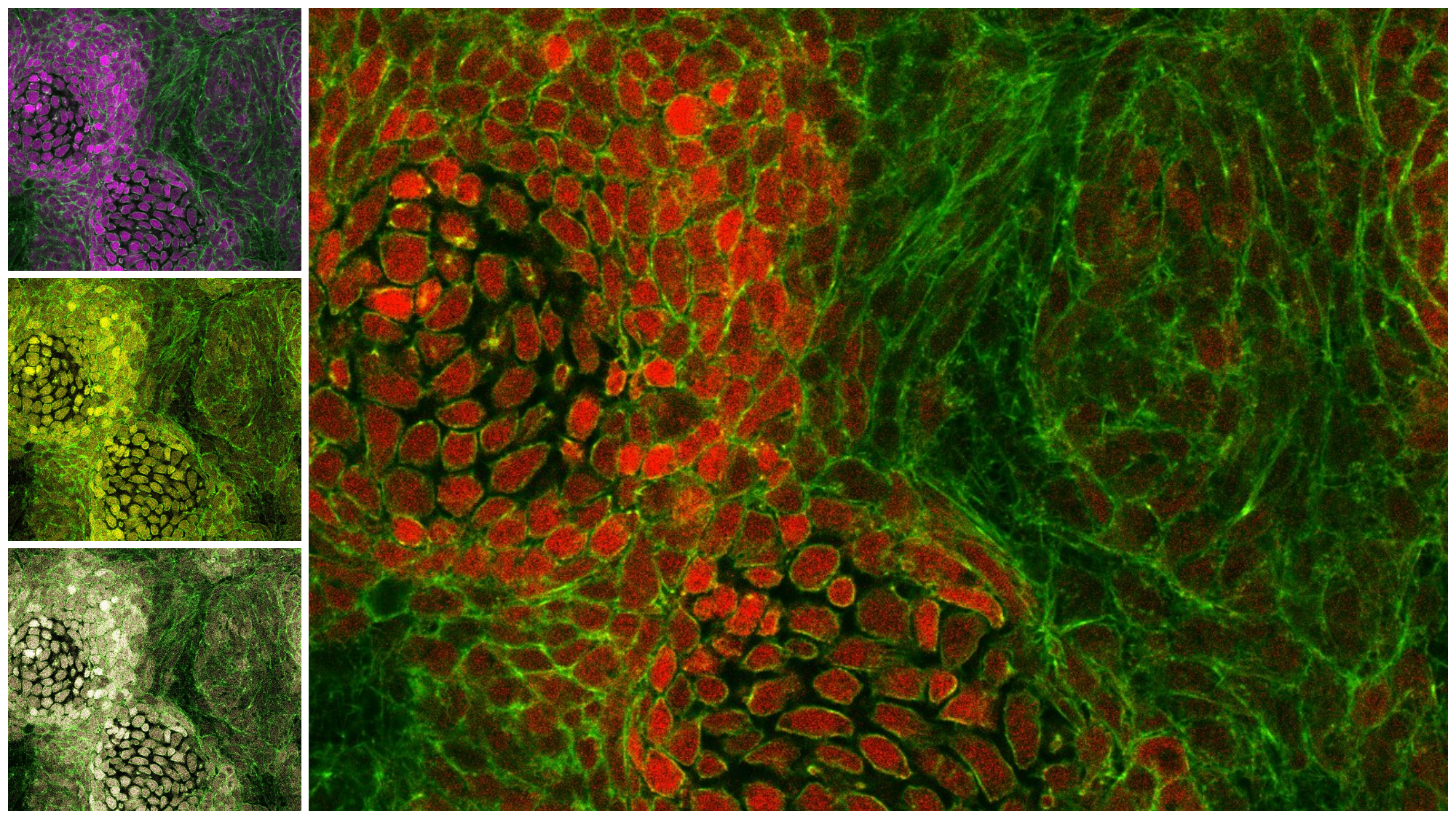
Heura Cardona, from the European Molecular Biology Laboratory – Barcelona (EMBL Barcelona), sends us this picture in which, to celebrate “Sant Jordi”, she has turned her cell culture into multi-colored roses!
The picture, taken with a Leica SP5 confocal microscope, shows a mesenchymal cell culture from mouse extremities at 11 days of development. These micromass cultures — quite dense cell cultures — are used as in vitro systems for drug screening and cell differentiation studies.
In this case, the researcher is studying how cells communicate and organize themselves and how cells that will become cartilage (the future bones of the extremities) and those that will become other tissues, rearrange themselves.
In red, we see the cells that express the transcription factor “Sox9“. These will become cartilage, while the cells that are not red (do not express Sox9) would correspond to the cells that will originate other tissues such as muscles, nerves, skin…
The cells are stained green with Phaloidin, which marks the actin of the cells — a component of the cytoskeleton of the cells.
Would you like to see your photo here? Please send us pictures related to science or the PRBB to ellipse@prbb.org.