For a long time, SARS-CoV-2, “the Coronavirus” in its most colloquial version, has been monopolizing covers, headlines and newsrounds around the world.
And since it is very difficult to understand that which we do not see, infinite representations of the virus have been created in all this time, from drawings for children to 3D models.
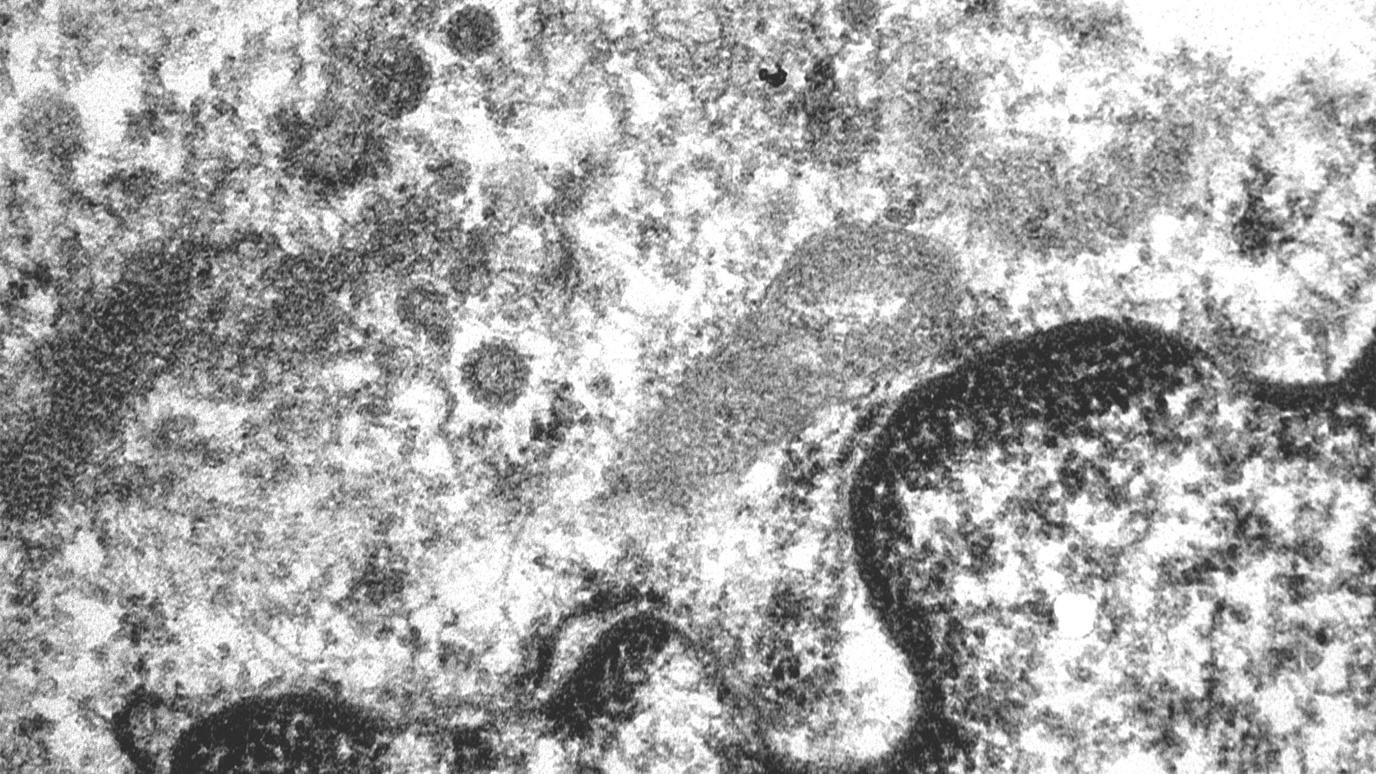
Here, thanks to the images and the explanation sent by doctor Josep Lloreta, head of the Pathological anatomy section of the Hospital del Mar and researcher at Hospital del Mar Medical Research Institute (IMIM), we can see the SARS-CoV-2 in the intestine of a COVID-19 patient:
“The image, taken by transmission electron microscopy with an original magnification x 34,000, shows various viral particles in a part of the nucleus and cytoplasm of one of the endothelial cells that line the blood vessels on the inside. It is mainly in the cytoplasm of these cells where SARS-CoV-2 reproduces and from where it colonizes the rest of the body. “
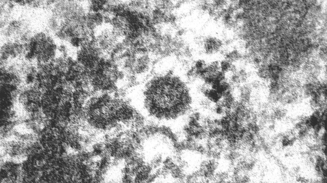
“In this second image we see SARS-CoV-2 in detail (original magnification x 130,000). Here we can appreciate its rounded appearance and the crown of anchoring pieces that gives it its name. This crown is the key element that the virus uses to hold on and enter the cells it wants to infect.”
Want to see your photo here? Send us your images related to science or life at the PRBB to ellipse@prbb.org.