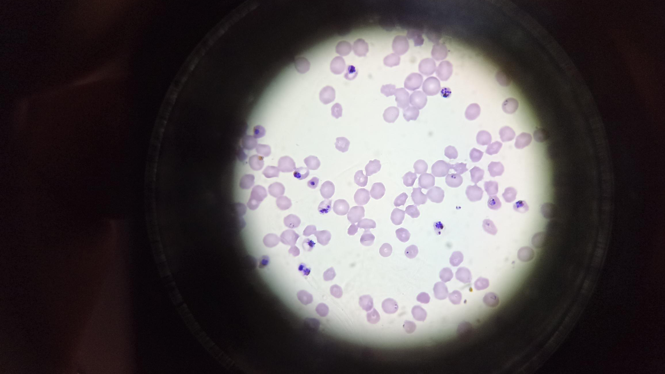
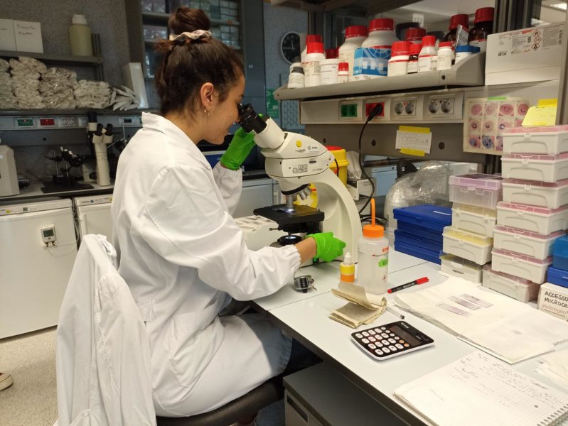
This ‘lab life’ picture was taken through an upright bench microscope at the laboratory lead by Maria Bernabeu, at the European Molecular Biology Laboratory – Barcelona (EMBL Barcelona).
As François Korbmacher, a postdoc in the lab who prepared this sample, tells us, the Bernabeu laboratory is studying the interaction of malaria parasites with in vitro blood vessels of the brain to better understand the pathogenic mechanisms of malaria infections. Malaria parasites can be grown in vitro by infecting red blood cells, which are received periodically from the blood bank.
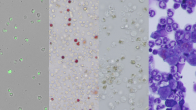
To observe parasite growth, the lab members fix and stain blood smears, one of the fastest and most commonly used methods for assessing parasite development that is widely used for diagnostics in endemic countries.
The human parasite infects the red blood cells in an asexual cycle of about 48 hours. Since it is mainly matured parasites that show pathologic effects, the laboratory is interested in parasites older than 24 hours after red blood cell infection. Therefore, the parasites are grown in a synchronized culture, for which the right timing needs to be assessed by blood smear stains. Therefore, the team needs to track the growth daily at the lab, as María Gestal Mato is doing in this picture below.
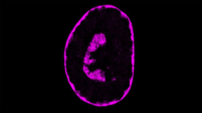
When the parasites are ready, they can be used for perfusion of in vitro bioengineered brain microvessels. This way, the laboratory dissects the parasite’s interaction with blood vessels and their impact on their health and function.