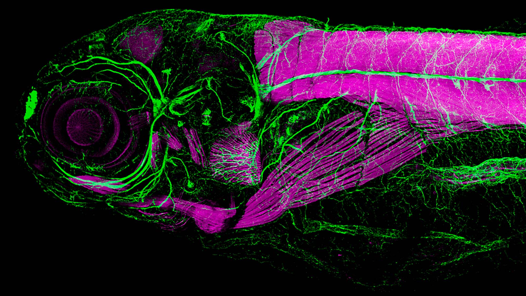
This image was sent to us by Nerea Montedeoca Vázquez, PhD student in the Morphogenesis and Signalling in Sensory Systems group of the Department of Medicine and Life Sciences at Pompeu Fabra University (MELIS-UPF), where they study inner ear development using zebrafish as a model. The image corresponds to an immunohistochemistry of the most anterior part of a 6dpf (six days post-fertilisation) zebrafish larva.
The magenta colour is due to the use of phalloidin coupled to a fluorophore (Alexa 568). This labelling shows F-actin, a type of molecule that is part of the cytoskeleton and is especially enriched in muscle cells.
In green we can see acetylated tubulin, an essential element in the formation of structures such as microtubules. This labelling allows us to see the axons of neurons and the structures of the ear.
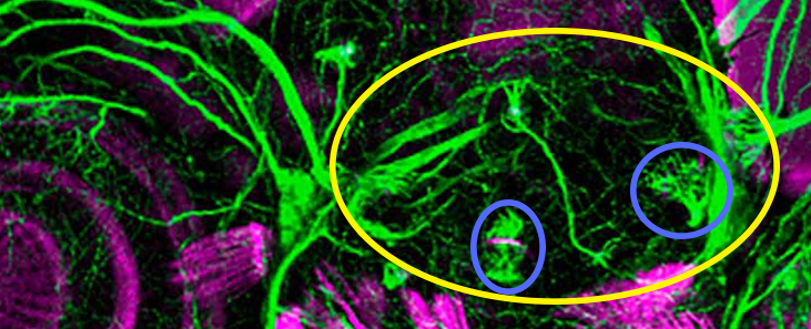
The eye of the larva can be easily seen thanks to its circular shape and magenta staining, and just to its right, marked in green, we see the ear (yellow circle in the previous image).
At this point in development, the ear consists of sensory structures, such as the maculae and cristae, marked in blue circles in the picture. These structures are made up of a group of ciliated cells that allow auditory and vestibular stimuli from the environment (sounds, information related to the position of the head in space, etc.) to be captured. To do this, these cells contain two types of extensions, called stereocilia and kinocilia, which are responsible for the mechanotransduction of stimuli.
And how does the mechanotransduction work? These prolongations are “linked” together and when they receive a stimulus, they move together in synchrony. This movement allows the opening of channels in the cell that allow an influx of ions to flow into the cell, which results in the information received being sent to the sensory neurons. These, in turn, send the information to the central nervous system where it is processed and interpreted.
Want to see your photo here? Send us images related to science or life in the PRBB to ellipse@prbb.org.