The microscope is one of the most widely used tools in biomedical research laboratories around the world. It allows us to see things that are invisible to the eye because they are too small for human vision, such as microorganisms, cells or tissues.
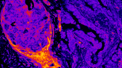
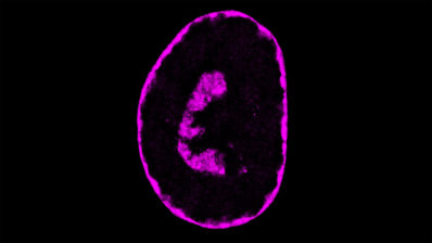
This image was taken in Thomas Surrey’s lab at the Centre for Genomic Regulation (CRG), where they are studying how the cell’s cytoskeleton organises itself during cell division, where it plays a key role in separating the chromosomes between the two resulting cells. The components of the cytoskeleton are proteins that are too small to be seen even under a microscope. To make them visible, they are tagged with fluorescent labels of different colours, which allow the different proteins to be located and their interactions to be seen using very powerful laser microscopes.
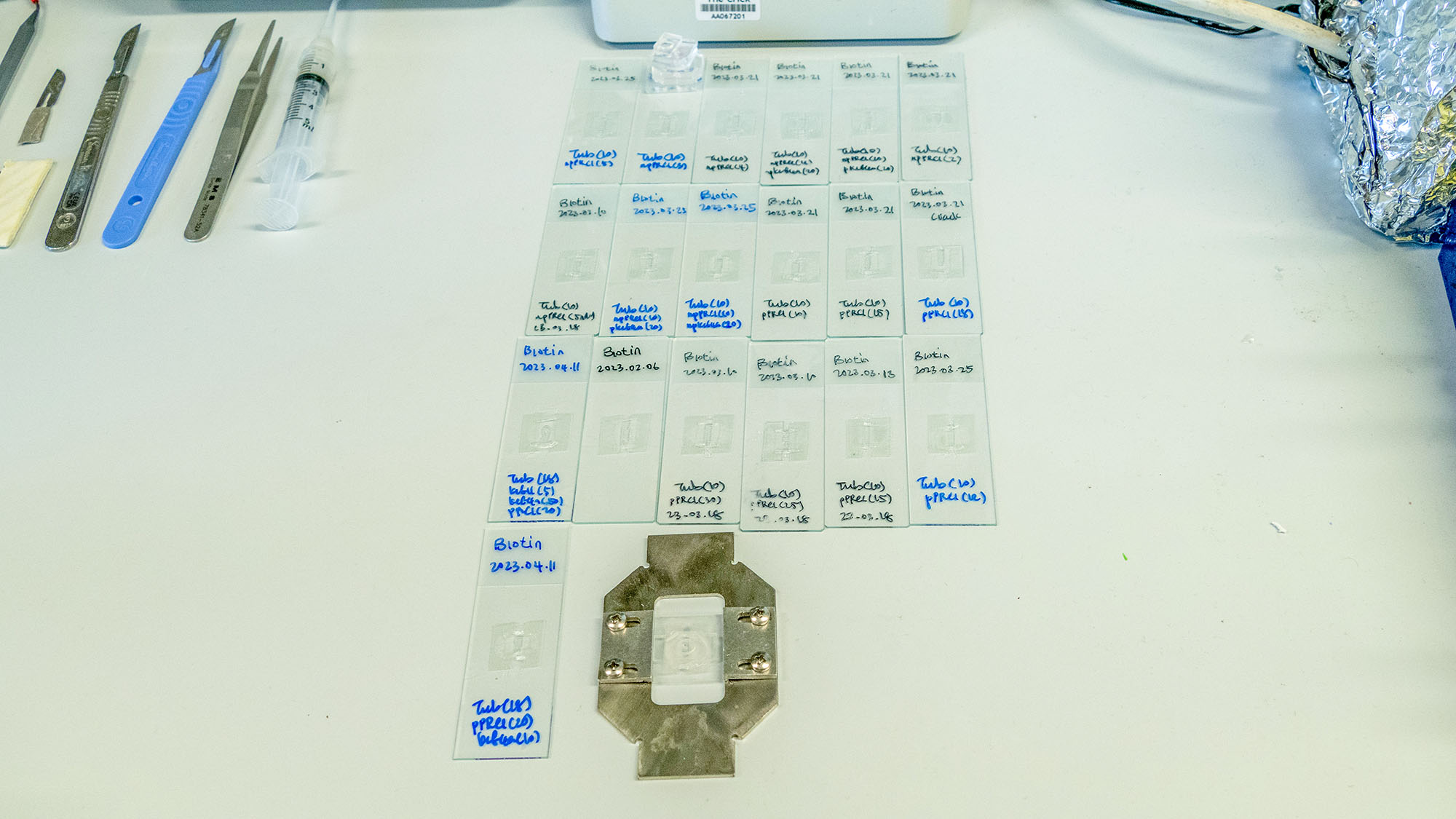
The glass slides needed for these experiments are very special and have to be made in the lab itself because there is no company that makes them. On top of the glass, they attach a small “fishbowl”, a micro-container of liquid, into which they can add the different proteins they want to see at different times and under different conditions.
On the slide they write which proteins they have added to each experiment (e.g. tubulin, that can be seen labelled in the image, is the main protein of the microtubules that form the cell cytoskeleton), as well as the different conditions that will be tested.
This way, the researcheres can see live how tubulin molecules move, how they join together to form microtubules, etc. In short, they observe one of the basic movements of life on a slide.