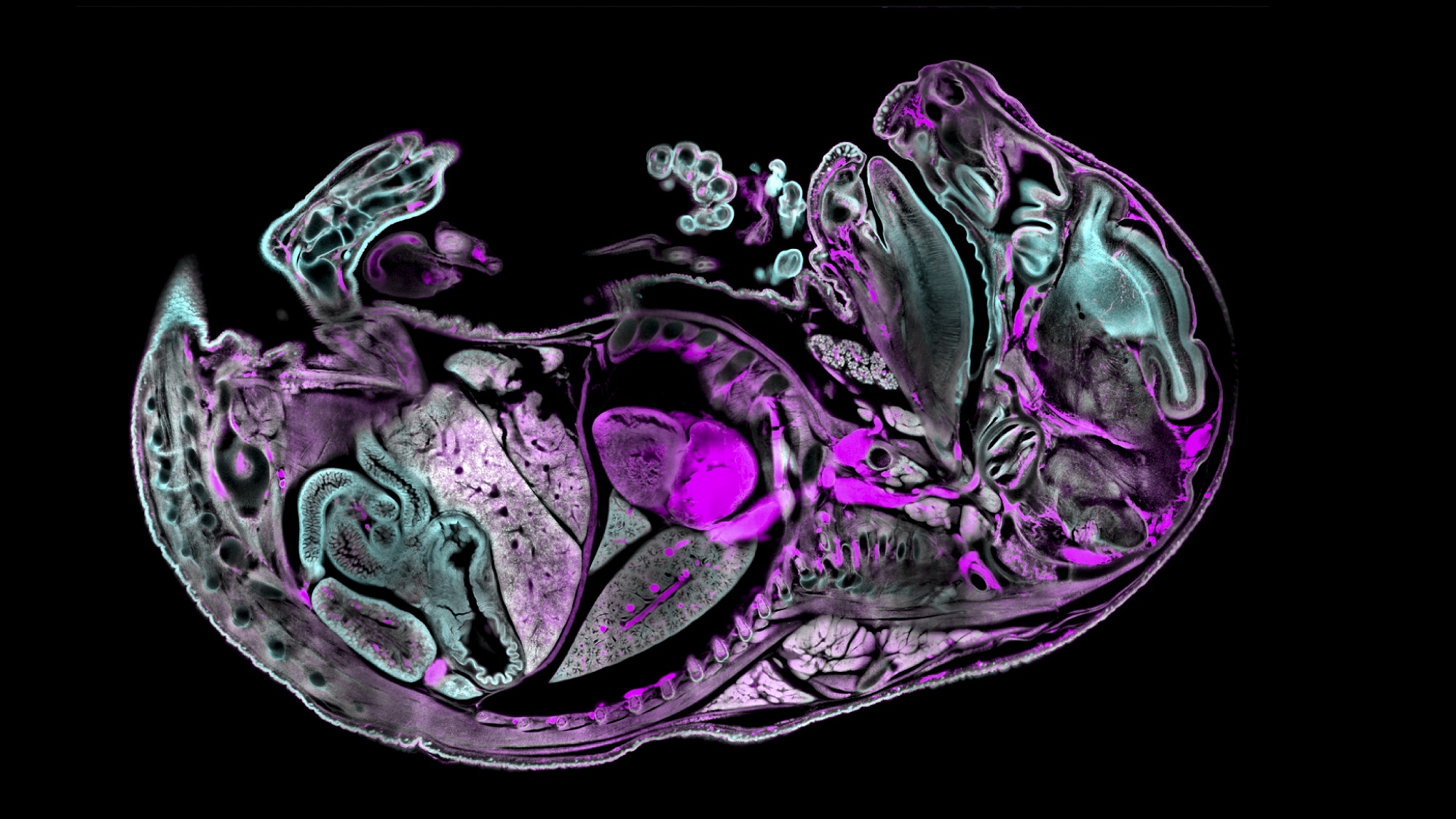
In this picture we can see a slice through 3D acquisition of a near newborn mouse, cleared with Benzyl Alcohol-Benzyl Benzoate (BABB), an organic solvent used to make samples transparent.
The mouse was stained with methylene blue (cyan) and is showing autofluorescence (magenta). The whole embryo can be seen, head to tail, with several anatomical structures visible – parts of the brain, spinal cord and digestive tract, as well as internal organs and limbs.
The sample was provided by Heura Cardona, from the Sharpe lab at European Molecular Biology Laboratory – Barcelona (EMBL Barcelona) Barcelona, and the labelling, clearing and imaging was done by Montserrat Coll Lladó from the Mesoscopic Imaging Facility (MIF).
Montse tells us more about it.
“At our facility, we received a new light sheet microscope, the LCS SPIM from Luxendo (Bruker), capable of imaging large optically cleared samples in 3D. We decided to push our limits by staining and clearing an exceptionally large specimen: a near newborn mouse over 1.5 cm in length.
The real challenge wasn’t the clearing itself, but ensuring that both the staining and optical transparency were consistent throughout the entire depth of the organism. To achieve this, several preparatory steps were carried out, including decalcification, pigment reduction (particularly in the eyes), and thorough permeabilization. Thanks to this approach, we obtained a uniform stain and clarity across the entire sample—quite a remarkable feat!”.
Indeed – congratulations to Montse and the whole team, and thanks for sharing this beautiful image!