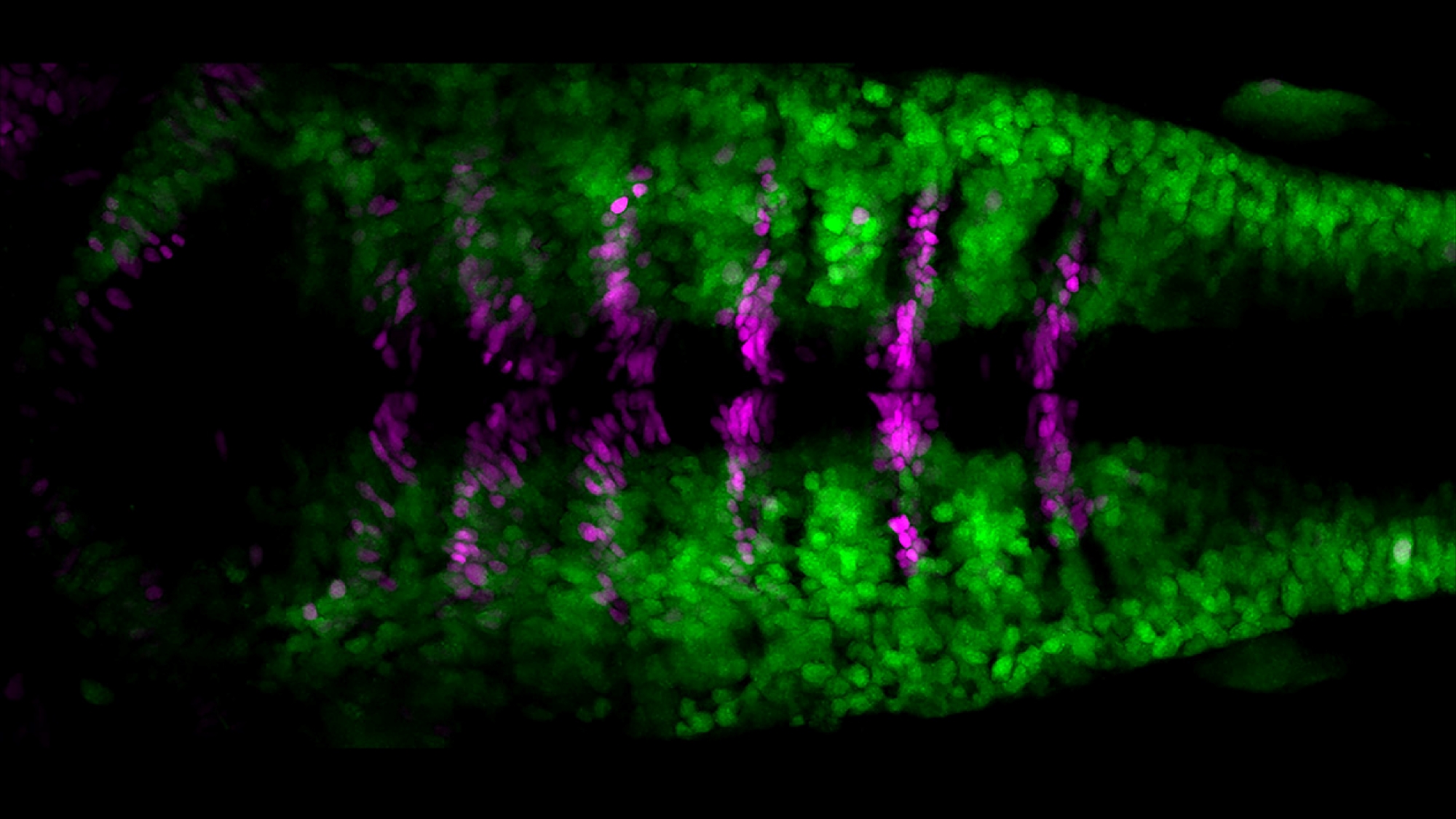
This photo from Cristina Pujades‘ laboratory at the Department of Medicine and Life Sciences, Pompeu Fabra University (MELIS-UPF) illustrates their latest study, in which they have observed, in the zebrafish model, the mechanisms that balance the number of brain stem cells and differentiated neurons during embryonic development.
The research group has studied the cell divisions that occur at the border between rhombomeres. These are each of the 7 segments into which the rhombencephalon or hindbrain, a conserved area in the brain of all vertebrates, is divided during embryonic development. In this way, they have been able to observe how brain stem cells follow a type of asymmetric division. That is, when a progenitor cell divides, it will form a differentiated neuron that can no longer divide and a new progenitor cell with the capacity to continue dividing. As seen in the photo, it is the cells at the border between rhombomeres that generate neurons (in green) while maintaining a proliferative reservoir of progenitor stem cells (in magenta).
Carolyn Engel-Pizcueta, author of the study, explains that “using CRISPR we have been able to insert a reporter gene and generate a transgenic zebrafish line where the cells of interest express a fluorescent marker so that we can observe and follow them over time”.
Thus, we have been able to elucidate the balance mechanisms that maintain the necessary proportion of stem cells and neurons in the brain. This is of great importance because, as Pujades points out, “during brain formation it is necessary to maintain the processes of proliferation and differentiation and to regulate them very precisely. Without this coordination, problems that cause neuronal disorders can arise”.
Hevia, CF, Engel-Pizcueta, C, Udina, F and Pujades C. The neurogenic fate of the hindbrain boundaries relies on Notch3-dependent asymmetric cell divisions. Cell Reports, 7 June 2022. DOI: https://doi.org/10.1016/j.celrep.2022.110915