In zebrafish, neurons in the peripheral nervous system and blood vessel cells talk to each other through dynamic protrusions called cytonemes or signaling filopodia. This communication helps regulate the proliferation and differentiation of neurons, according to a trio of researchers led by Berta Alsina, principal investigator of the Morphogenesis and Signaling in Sensory Systems group at Department of Experimental and Health Sciences, Pompeu Fabra University (DCEXS-UPF).
Decisions, decisions, decisions
Once born, the neurons in the peripheral nervous system must decide between:
- stay quiescent (stay latent, asleep, until it is necessary to reactivate them)
- proliferate (multiply, to increase the number of neurons)
- differentiate (become specialized neurons, for example auditory, nociceptive or Purkinje neurons)
It is important that there is a balance between the neurons that make each decision, in order to have the right number of neurons at the right time. Otherwise, problems can arise, which in the case of ear neurons could lead to deafness or vertigo, for example.
But cells are not pre-programmed to follow one of these paths by default; rather, the decision depends on communication with the other cells in their environment, which form their niche.
The blood’s role
It has recently been known that, at the level of the central nervous system, blood vessels communicate with neurons at the physical level and regulate the pluripotency of stem cells.
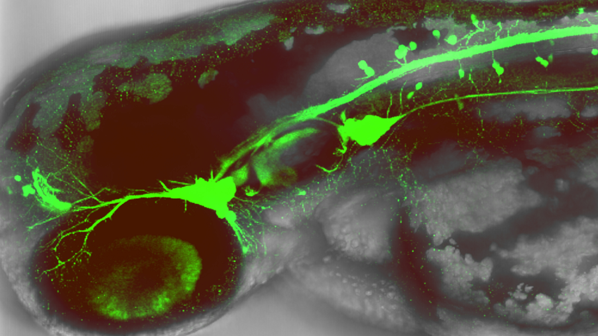
“People had seen that neurons envelop the vessels, that they physically touch each other. But what we have seen, observing it live, in real time, is that these contacts are made through filopodia and are dynamic, which allows a very fine regulation in terms of time and space, because they can decide which cell they touch and when”, explains Laura Taberner. The study is the result of his doctoral thesis.
“Neuronal precursors and blood vessels contact each other dynamically through filopodia, allowing for very fine regulation in terms of time and space”
Laura Taberner
The team led by Berta Alsina has seen these connections, for the first time, in the peripheral nervous system, specifically in the ear of the zebrafish. The research team used zebrafish embryos with fluorescent markers on both endothelial cells (those that make up blood vessels) and neuronal precursors in the ear.
The scientists have been able to determine that there are two types of contact between blood and neurons, which take place at different times.
- Between 24 and 36 hours of development, endothelial cells form cytonemes that touch some of the ear’s neuronal precursors, causing them to stop proliferating and remain quiescent.
- Between 48 and 60 hours, the cytonemes or filopodia disappear, but the blood cells again play an important role in bringing oxygen to the neuronal precursors, which produces a metabolic change that causes them to go from proliferating to differentiating.
“I had a great time developing this new project in the laboratory, I learned a lot and we provided knowledge that is relevant,” says Taberner, who after the thesis also did a master’s degree in education and now hopes to take a turn in her career to teach high school students.
Hear Laura Taberner talking about this project (video in Catalan).
L Taberner, A Bañón, B Alsina. Sensory neuroblast quiescence depends on vascular cytoneme contacts and sensory neuronal differentiation requires initiation of blood flow. Cell Reports, July 2020. https://doi.org/10.1016/j.celrep.2020.107903.