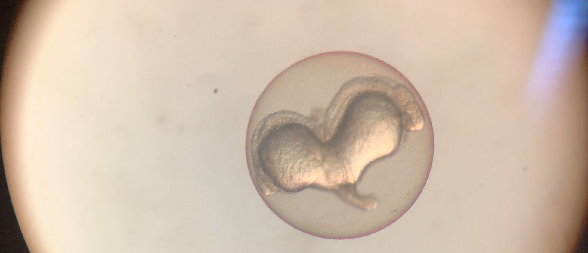
This beautiful image sent by Carla Belmonte Mateos, a researcher at the Department of Experimental and Health Sciences, Pompeu Fabra University (DCEXS-UPF) shows us an impressive example of siamese zebrafish embryos.
Carla works in Cristina Pujades‘ lab, where they study the development of the embryonic central nervous system (CNS) with the focus on the hindbrain, as it is one the most conserved embryonic structures of the brain.
The transgenic line of zebrafish observed in the picture expresses the fluorescent protein mCherry under the control of different krx20 regulatory elements, a transcription factor that is expressed in very specific regions of the embryonic hindbrain. This is a fantastic tool to use as a tissue landmark, as it helps you contextualize a population of interest within the whole structure.
Carla tells us what she was doing when she came accross this image: “I crossed adult fish overnight and collected embryos the next morning, as I do every other day to start an experiment. After 24 hours post fertilization, I checked the stage and health of the embryos. To my surprise, among a 200 spawn, I observed these siamese embryos creating a heart shape. I had previously seen an embryo with two heads, but I had never observed two hearts in one!”.
Want to see your photo here? Send us your images related to science or life at the PRBB to ellipse@prbb.org.