The brain, a vital organ for the proper functioning of the human body, is one of the main objects of study in the biomedical research.
Although we have not yet figured out how this organ works in every single situation and stage of life, much of the current knowledge has been obtained through the model animals used in research, and zebrafish is a good example.
The research group led by Cristina Pujades at the Department of Experimental and Health Sciences of the Pompeu Fabra University (DCEXS-UPF) studies the brain of this model organism. Recently, the group has proved that boundary cells located at the posterior brain of zebrafish transduce the mechanical stimuli into biological behaviours that will control cell fate during brain formation. Specifically, in response to mechanical stimuli, the progenitor cells decide whether they keep on dividing or they differentiate into neurons.
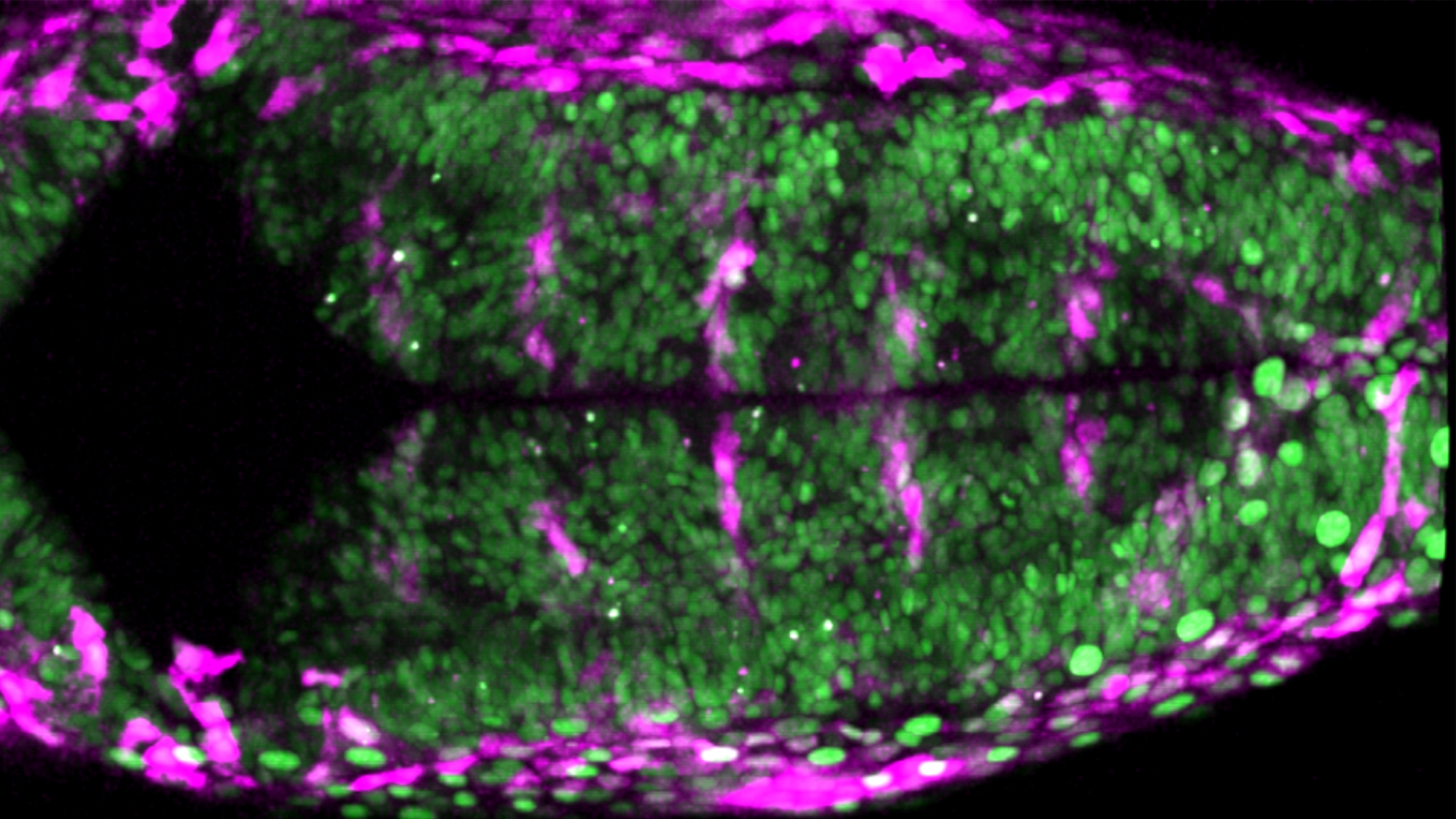
In the image above we can see a dorsal vision of a zebrafish posterior brain. The picture, obtained from the study performed by the Pujades lab, shows us:
- Cell nuclei in green.
- Cells responding to mechanical signaling in magenta.
This finding leads the research group to conclude that “mechanical forces generated during embryogenesis controle the balance between neurons proliferation and differentiation“, according to Pujades.
Would you like to see your photo here? Please send us pictures related to science or the PRBB to ellipse@prbb.org.
Voltes A, Hevia CF, Engel-Pizcueta C, Dingare C, Calzolari S, Terriente J, Norden C, Lecaudey S, Pujades C. Yap/Taz-TEAD activity links mechanical cues to specific to cell progenitor behavior during hindbrain segmentation. Development 146: dev176735, July 2019. doi: 10.1242/dev.176735.