A study led by the Centre for Genomic Regulation (CRG) has used super-resolution microscopy to obtain the most precise images of transcription to date. They have collaborated with the Institute of Photonic Sciences (ICFO), the Bioland Laboratory of the Guangzhou Institute of Biomedicine and Health (GIBH), in China, and the University of Pennsylvania.
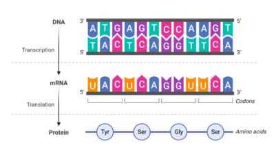
Transcription is the transfer of information in the form of DNA to RNA on its way to the formation of proteins. With the technique used, the position of individual molecules can be obtained, which, combined with computational analysis, has given more information about how RNA is arranged in a human cell during this process.
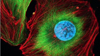
The results, with a resolution up to 10 times higher than that achieved with fluorescence microscopy, show with nanometric precision that the newly transcribed RNA molecules are organised into small domains. Furthermore, the researchers have observed how these nanodomains physically interact with chromatin and the rest of the transcription machinery. Thus, it has been shown that “transcription is a finer, more precise and regulated process than previously thought”, according to Álvaro Castells, a CRG alumni and current GIBH researcher.
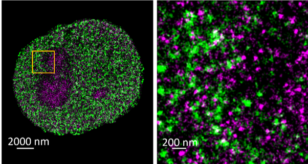
The use of this microscopy technique has opened the door to a better understanding of how transcription works and how molecules are organised during this process. As Pia Cosma, principal investigator at the CRG and leader of the study, says, “this study has helped us to visualise and locate the emerging RNAs with unprecedented precision“, adding that “in the future, this type of analysis can be used to understand gene expression with greater precision”.
Nucleic Acids Research, Volume 50, Issue 1, 11 January 2022, Pages 175–190, https://doi.org/10.1093/nar/gkab1215


