There is no better way to find the answer to a problem than asking questions from different points of view or, in this case, from different microscopes: different concepts, different lenses, and clearly different results.
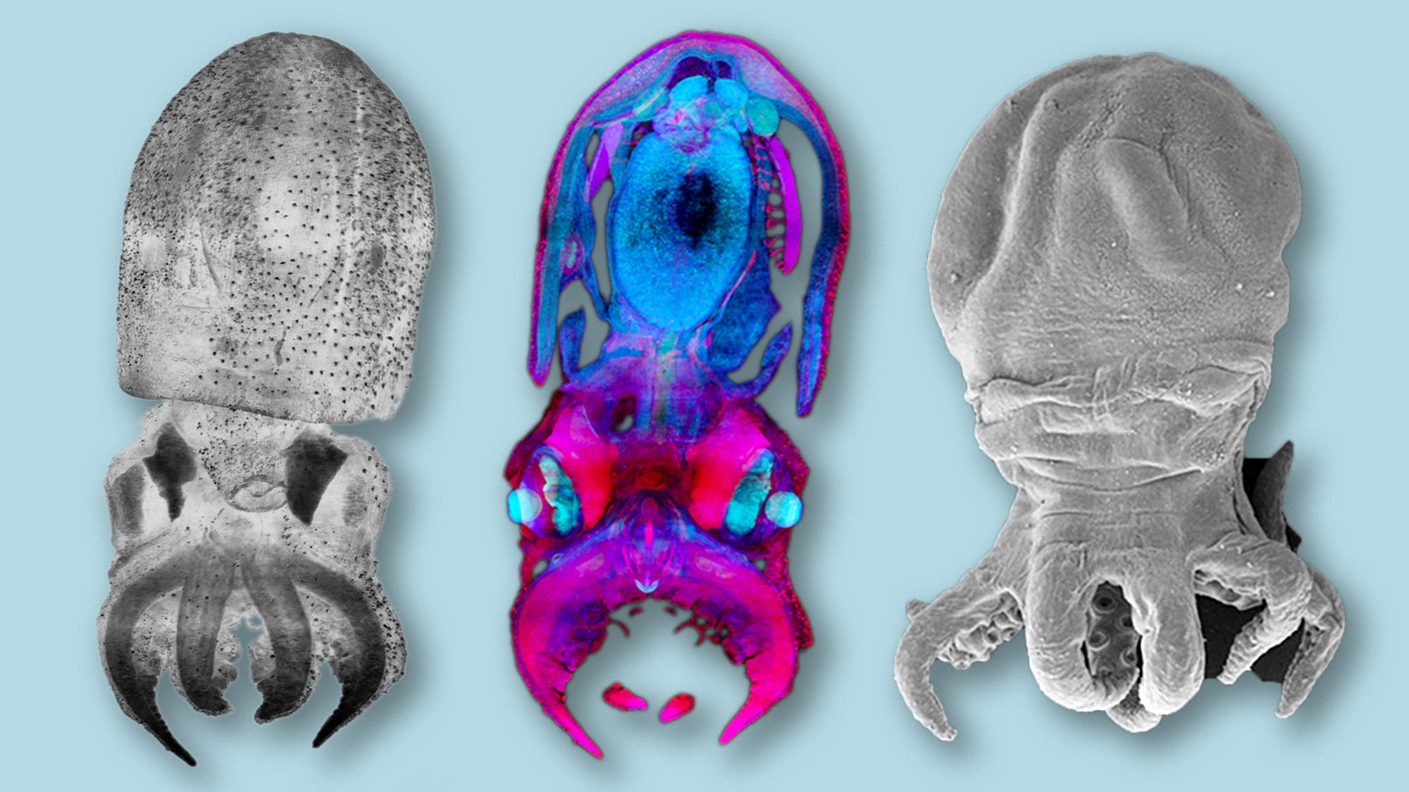
In the image above – from a study on the octopus Kölliker’s organs (KO) – we can observe three images taken with two different microscopy techniques.
The first technique is light sheet microscope, or Selective Plane Illumination Microscopy (SPIM), which allows to observe a complete sample without doing histological sections. This technique was applied at the Mesoscopic Imaging Facility of the European Molecular Biology Laboratory – Barcelona (EMBL Barcelona) to samples of an Octopus vulgaris or common octopus, to obtain the first two images.
The last image on the right shows the same octopus observed with a scanning electron microscope (SEM) in which the surface of the sample is scanned, but not its interior.
To learn more about the Mesoscopic Image Service check this article and don’t miss the video below (in Catalan) in which Montse Coll, responsible of the photographs, explains the different types of microscopes, their operation and the procedure to obtain these fantastic images.
Want to see your photo here? Send us your images related to science or life at the PRBB to ellipse@prbb.org.
Villanueva R., et al. Born With Bristles: New Insights on the Kölliker’s Organs of Octopus Skin. Frontiers in Marine Science, 10 May 2021


Each technique offers a unique window into the microscopic realm. For example, electron microscopy delves into the ultrastructural level, revealing organelles and their internal landscapes, while live-cell imaging captures dynamic processes like cell division and protein trafficking.