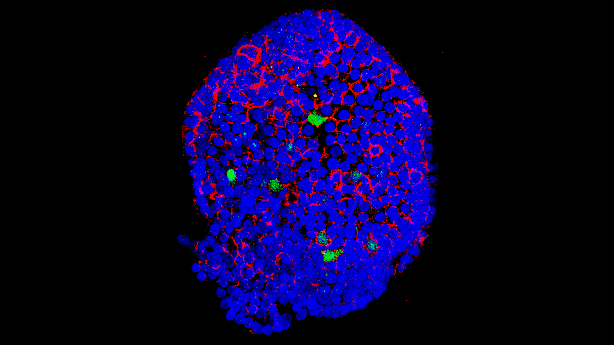
This picture, taken with a confocal microscope, shows a whole organoid derived from human lung tissue. The blue colour (DAPI) is a nuclear stain that labels the DNA; the red stain (phalloidin) binds to actin and it indicates the cell membrane; and the green stain (UCHL1) is a marker of pulmonary neuroendocrine cells.
The image was taken by the laboratory lead by Talya Dayton, a new PI at the European Molecular Biology Laboratory – Barcelona (EMBL Barcelona). They study the pulmonary neuroendocrine cells (the ones marked in green), which secrete hormones and neuropeptides, to understand their differentiation and plasticity, and also to understand how neuroendocrine cancer develops.
The lab generates these organoids from tissue stem cells (in this case, lung stem cells), and they also look at them live, as in the picture below:
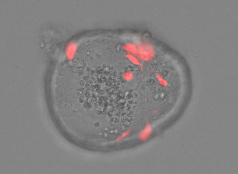
In this case, the image shows the overlay of the fluorescence – where the pulmonary neuroendocrine cells are now seen in red because they express a fluorescent neuroendocrine reporter – and the brightfield image of the organoids.
Want to see your photo here? Send us images related to science or life in the PRBB to ellipse@prbb.org.