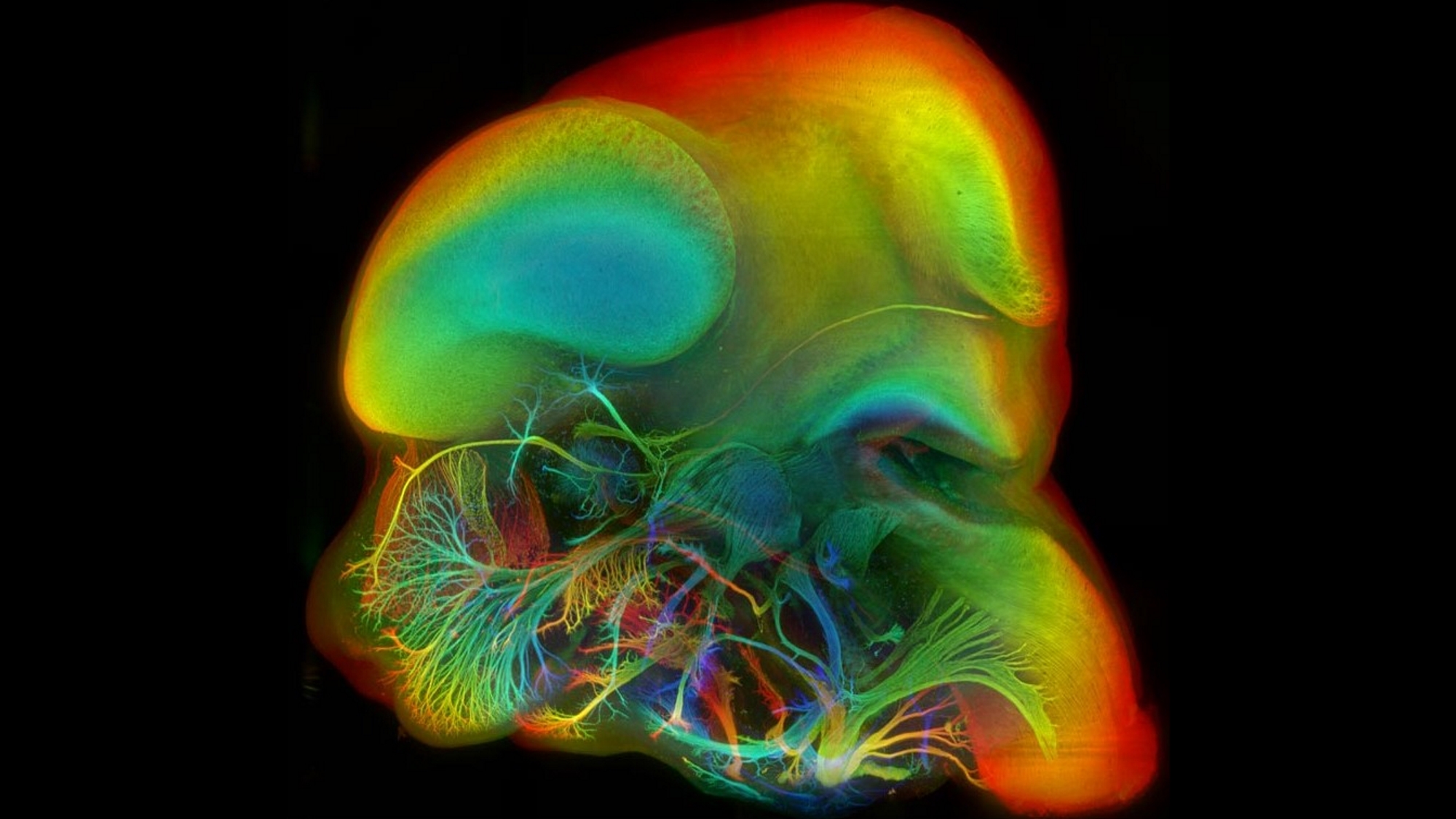
The head of an embryonic mouse about 12 days post-fertilisation was fluorescently labelled to enable the visualisation of neural precursor tissue, chemically treated to make it optically transparent, and scanned in the Sharpe lab at the Centre for Genomic Regulation (CRG) (now at the European Molecular Biology Laboratory – Barcelona (EMBL)) using a SPIM (Selective Plane Illumination Microscope, or Light Sheet Microscope) to generate a 3D virtual reconstruction.
In this type of microscope, a thin sheet of laser light illuminates a single plane in the sample, so that only the part that is in focus in the camera fluoresces. The sample is then scanned using the light sheet to build up a full 3D data set, plane by plane. Using this 3D data, we generated a maximum-value projection through the left half of the embryo’s head, with false colour to encode the depth in the sample (blue=more superficial, red=deeper). Sample courtesy of Glenda Comai, Pasteur Institute, Paris.
Would you like to see your photo here? Please send us pictures related to science or the PRBB to ellipse@prbb.org.