Just like Harry Potter and the rest of the Hogwards quidditch seekers’, research staff looking through the microscope for results, are occasionally lucky enough to capture images as precious as the golden snitch.
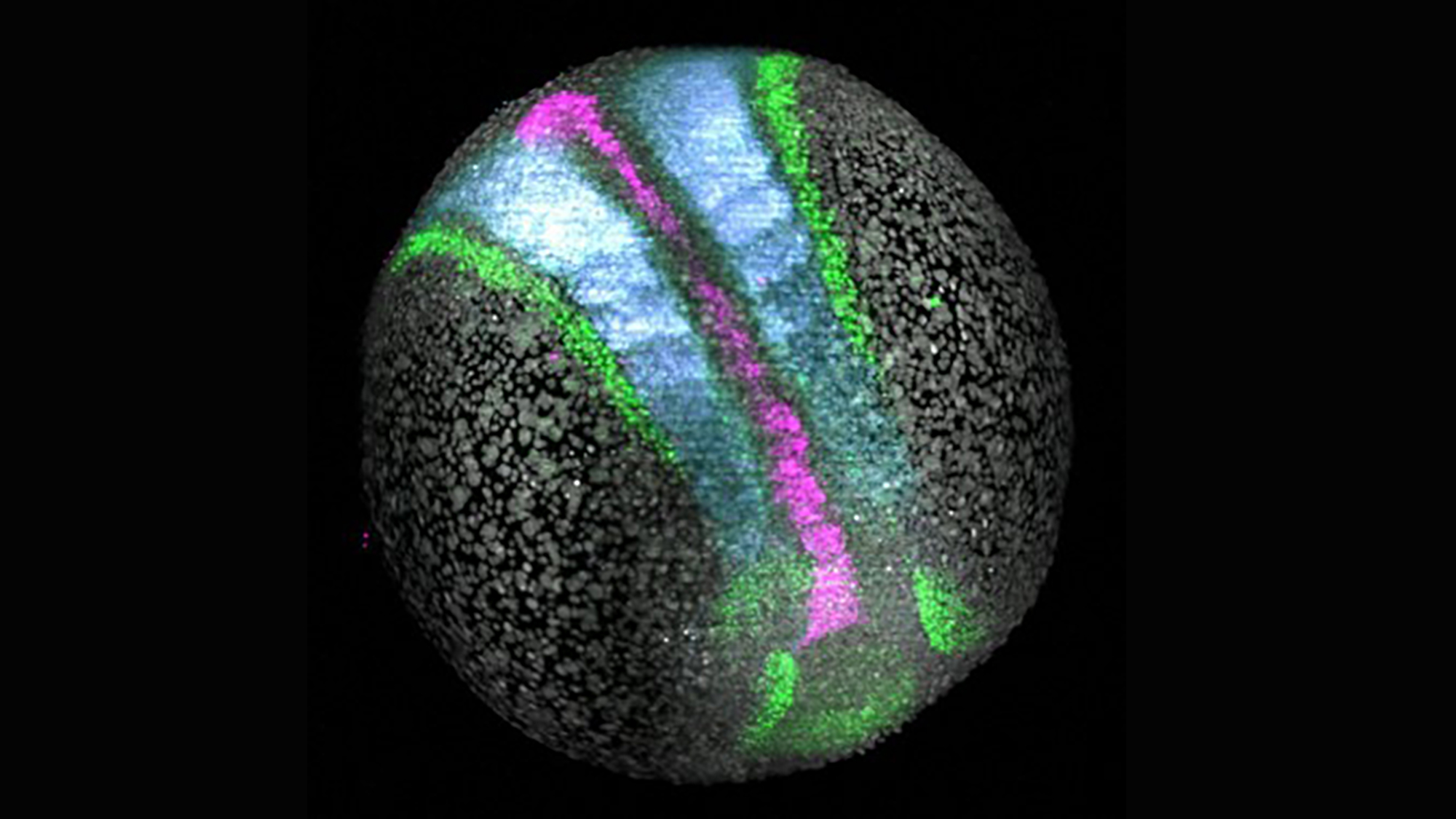
This is what happened to Nicola Gritti, now image analysis specialist in the mesoscopic imaging facility at the European Molecular Biology Laboratory – Barcelona (EMBL Barcelona), while watching zebrafish embryos develop.
This fish, which is widely used in research, has a rapid development. Therefore, ten hours after fertilization, cells that begin to differentiate and express genes characteristic of a germ layer or tissue can already be seen.
During his postdoc in the Trivedi lab at EMBL Barcelona, Nicola used hybridization chain reaction to identify the cells expressing Pax2a, which will become part of the nervous system (green); those expressing Meox1 that will become somites (blue) and those expressing Foxa2 that will end up forming part of the endoderm (lilac).
All this ended up drawing this image that reminds us of the desired prize by quidditch seekers.
Would you like to see your photo here? Send images related to science or life to the PRBB to ellipse@prbb.org.