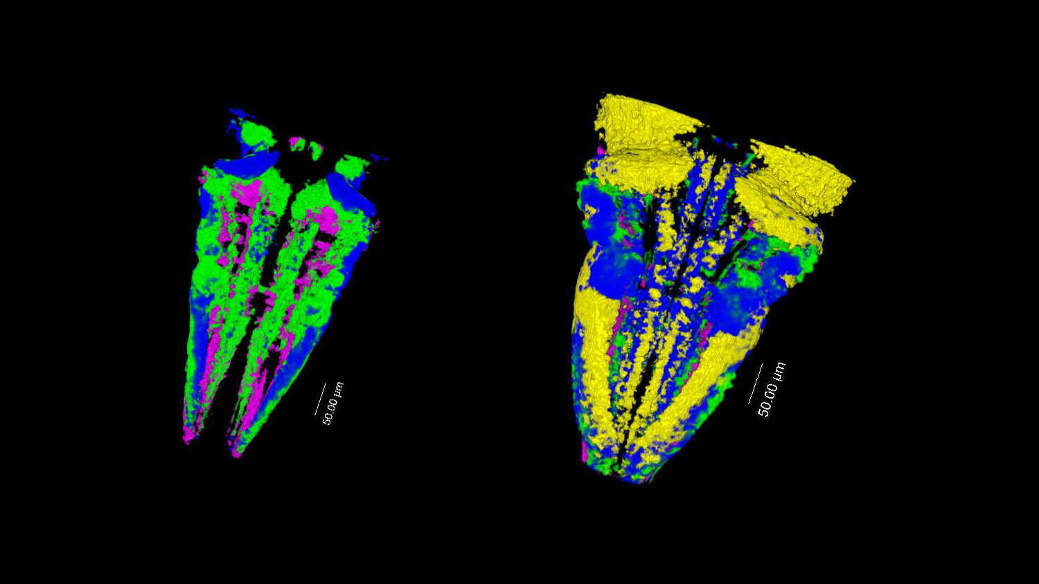
This image shows an image of the 3D digital atlas of the vertebrate brain obtained thanks to the DAMAKER tool. It comes from the Neurodevelopmental Dynamics Group of the Department of Medicine and Life Sciences, Universitat Pompeu Fabra (MELIS-UPF), led by Cristina Pujades.
The image presents a map of the birth of glutamatergic (excitatory) neurons in the zebrafish hindbrain. This is the part of the vertebrate brain that is responsible for basic functions such as swallowing and breathing. On the left we observe these neurons at 48h of life and on the right at 72h after their differentiation.
“The microscopy and image analysis techniques we have today are impressive and allow us to see in real time the movement of cells and the formation of many biological structures”
Cristina Pujades, MELIS-UPF
3D temporal map of different neuronal populations.
DAMAKER is a tool that allows to obtain digital atlases of 3D models that integrate spatial and temporal data. The program, created by the UPF team in collaboration with the European Molecular Biology Laboratory – Barcelona (EMBL Barcelona), combines in vivo images of cells marked with fluorescent markers that are tracked throughout their development and analyzed using freely available algorithms. This means that the program can be used by anyone to build an atlas of any cellular tissue as long as specific markers are used for what they want to see.
Thanks to the program, Cristina’s team has produced a 3D atlas of the zebrafish hindbrain. In addition to knowing when neurons differentiate, they have shown that the place and time of birth of these cells mark their final destination.
This study presents the first mapping of the vertebrate brain, and the digital 3D atlas generator could be a good tool to get to know the life history of cells during morphogenesis, the process of tissue formation. Also, thanks to zebrafish models, human avatars with mutations could be created to study diseases. “Instead of looking for neuronal affectations as if they were a needle in a haystack, we can go to a certain stage, look at the distribution of neurons and, by comparing it with the atlas, we can know when the neuronal defect occurred”, Cristina points out.
Do you want to see your photo here? Send us images related to science or life in the PRBB to ellipse@prbb.org.
Matthias Blanc, Giovanni Dalmasso, Frederic Udina, Cristina Pujades. A dynamic and expandable Digital 3D-Atlas MAKER for monitoring the temporal changes in tissue growth during hindbrain morphogenesis. eLife 2022. DOI: 10.7554/eLife.78300.