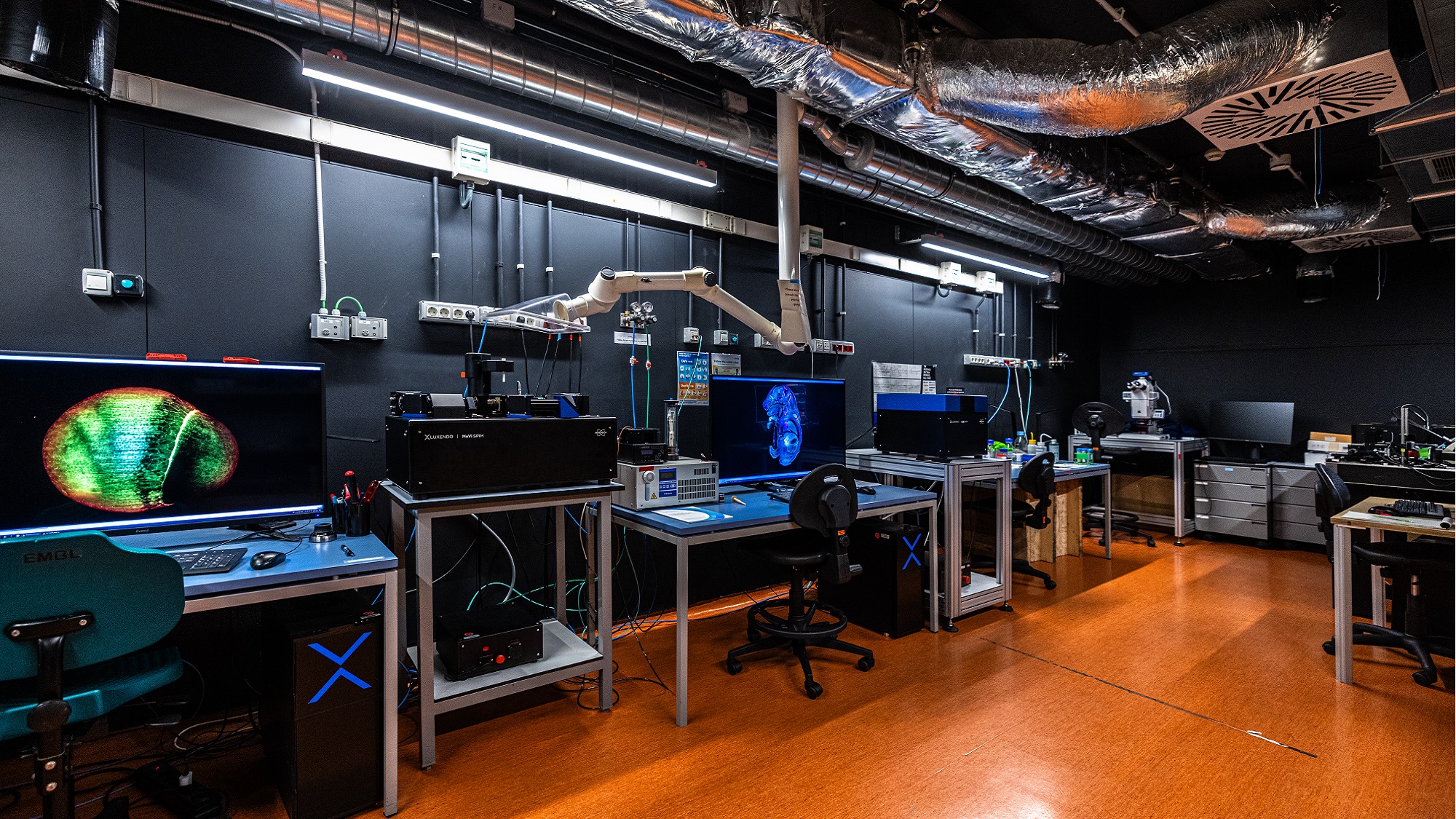
There’s more than meets the eye at the Barcelona Biomedical Research Park (PRBB) – figuratively and literally. Out of sight, on the -1 floor of the building, one can find not only one of the most complex and automated animal facilities in Europe, but also two state-of-the-art imaging facilities: the Mesoscopy Imaging Facility (MIF) from the European Molecular Biology Laboratory – Barcelona (EMBL Barcelona) and the Advanced Light Microscopy Unit (ALMU) from the Centre for Genomic Regulation (CRG), located in the same space. “For the last 6 years, we have offered complementary services to the ALMU; they offer confocal and super-resolution scale microscopy. We offer mesoscopy”, states Gopi Shah, Manager of Strategic Imaging Initiatives at the MIF, EMBL Barcelona.
Mesoscopy: the in-between
Microscopy deals with very small things: samples at the nano to micron level sizes, such as tissue sections, single cells, subcellular structures, organelles etc. On the other side of the scale are X-rays, MRI, CT scans and such; technologies which we more often associate with hospitals, and which are useful for looking at the human patient level. Mesoscopy represents the scale in between – and it adds volume.
The usual suspects that are imaged with mesoscopy systems are either whole embryos of model organisms (fly embryos, fish embryos, mouse embryos) or whole organs, such as brain, lungs, kidneys, spleen, ovaries etc. from adult model organisms.
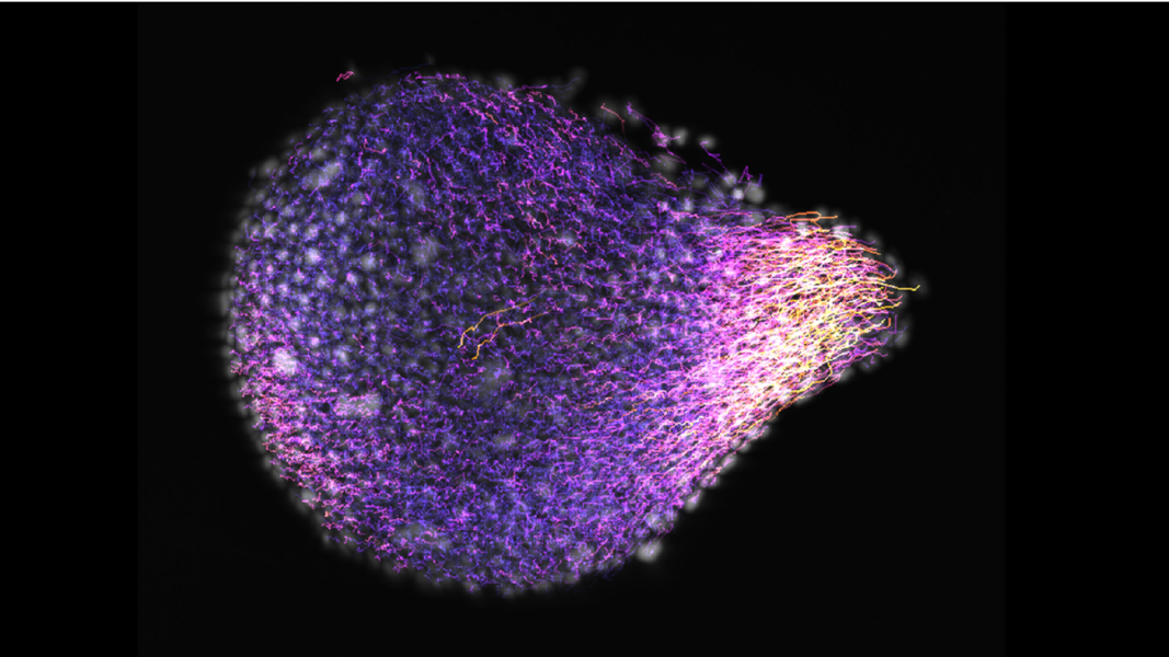
The kind of sizes you can see with mesoscopy go from a few hundred microns to a few centimetres – with some exceptions. “We’ve been able to visualize the growth and development of live organoids and other in vitro models that have been developed in recent years. These are a few hundred microns in size, so on the smaller side for your usual mesoscopy, but it is difficult to look inside them with other microscopes (confocal, super-resolution) as they are 3D and densely packed samples. Our systems are useful for them as they enable us to view samples from multiple directions and reconstruct a full 3D volume. Also, researchers from the Marine Science Institute (Institut de Ciències del Mar (ICM – CSIC) next door bring us samples that are 20-50microns in size, and we still manage to deal with them with some of our systems”, says Gopi.
The MIF is open to everyone, since it receives core funding from the European Molecular Biology Laboratory, Europe’s Life Sciences Laboratory. “We are happy to work with very diverse users and institutions – from EMBL colleagues, to PRBB neighbours, to labs/hospitals/industry partners in Barcelona and beyond. Our main aim is to provide cutting-edge service to the scientific community”, says the MIF manager.
Our main aim is to provide cutting-edge service to the scientific community, wherever they come from.
Gopi Shah, Manager of Strategic Imaging Initiatives, MIF, EMBL Barcelona
Regarding the cost, she adds: “So far, we have not been charging for using the facility. Every new system needs a lot of testing, and users have helped us with that. Therefore, we are currently running the facility with a collaborative approach and in future our services may be charged at affordable rates. We are committed to building collaborations and offering cutting-edge imaging service to as many labs as possible”.
Different microscopes and different techniques
Light-sheet fluorescence microscopy (LSFM, also called Selective Plane Illumination Microscopy or SPIM) is the main type of imaging done at the facility. They have 2 homemade set ups and 3 commercial set ups, each with different applications. The homemade ones can be customized for things that cannot be done with commercial ones. They look at large samples such as a whole mouse embryo or even an adult mouse brain at single cell resolution. “We also use these systems for imaging live organoids – you can do imaging for days and the organoids are comfy there!”, explains Gopi.
Optical projection tomography (OPT), another of the techniques they offer, uses a light source and a camera, putting the sample in between. It works by rotating your sample 1 degree at a time and taking a picture. Like that, you end up with 360 images and you can reconstruct the 3D object in the same way that an x-ray CT system in a clinic allows doctors to make a 3D reconstruction of your bones. These are useful for larger samples, and the facility owns two OPT machines, one commercial and one homemade.
A third type of technology they have at the facility is holotomography: it enables live imaging of cell cultures in a dish and flat cell sheets which are label free (no need for dyes or fluorescent labels, as it uses inherent contrast that comes from the fact that each compartment in a cell has a different refractive index) and you can see the organelles inside the cells. “The one limitation is that it can only capture a depth of 30 microns”.
All these imaging systems allow different ways of visualizing the samples, be it live or treated.
“We can do Tissue clearing, which is a technique to look deeper into a sample; you apply chemicals so that it becomes transparent and uniform in its refractive index throughout”, explains Gopi, who adds this is useful to see a whole mouse organ or even a whole adult mouse. “You can dissect an organ, perform labelling, do the tissue clearing with the chemical reagents and look at it with light-sheet microscopy or OPT”. Holotomography is the only one that cannot be used for cleared samples.
Apart from their specialized microscopes and techniques, they have some more usual equipment, such as an epifluorescence microscope to run a time-lapse, or to check if the labelling has worked properly, a tissue culture hood in case some small manipulation needs to take place, and a wet lab area where users can prepare their samples if needed.
A step-by-step process
Usually, when a user is interested in using the facility there’s a 3-step process that is followed:
- Meeting/consultation where researchers explain what they want to achieve. The facility staff show them around what they have and discuss what’s possible and what’s best suited for the task.
- The users receive training (2-3 sessions) and then they can register, book and use the instruments. They are also advised on sample preparation: how to keep the live samples alive, how to do the sample mounting and tissue clearing, etc.
- Image processing and data visualization and analysis: the MIF has a person dedicated to image processing to help users to handle the data, and who can also write custom codes to help with image analysis.
The unit generates tonnes of images. “Light-sheet is really fast – it can record a beating heart of a zebrafish, a 3D structure that beats 2 times/second”, says Gopi. According to her, if all the facility’s instruments are being used, that’s several terabytes of data per day. That’s why they offer a temporary data storage space where users can keep the data for downstream processing and analysis.
In-house expertise
The MIF is an interdisciplinary, international team of 5 people, with research backgrounds in biology, engineering and physics.
“We have 2 optical engineers, 2 biologists and an image analyst who enable the different steps of an imaging experiment. But everyone, whatever their original background, has specialized in imaging”, explains Gopi, who has the expertise in live sample imaging.
Montserrat Coll Lladó is the expert in tissue clearing. They also have two optical engineers who build the microscopes. Jim Swoger, who is also the head of the facility and Spyridon Bakas, who is currently working on developing a new bench-top system and to customize the commercial systems – e.g. if an experiment needs a new sample holder, or someone needs any physical tool, he makes them in the microfablab, located on the -2 floor of the PRBB. For the homemade equipment, sometimes the parts are commercial and sometimes designed specifically in a workshop or 3D printed. They obviously need specialized conditions to develop these systems, and they have big antivibration tables in the facility for their development.
Nicola Gritti is an expert in image data handling, processing, and analysis. He helps users with training and customization of image processing pipelines. Together the team offers end-to-end support to facility users.
Expansion microscopy
The team has lately beenexperimenting to try and see smaller things. There are two main ways to achieve this:
- The optical way. Normally one cannot see two objects that are closer than 200nm, but with super-resolution microscopy techniques one can go down to 20nm. Such super-resolution microscopy is available at the CRG’s ALMU unit, which shares space with the MIF.
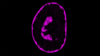
- By expanding the samples isotropically. That means to separate all the structures and molecules in a sample in a way that the relationship between them stays the same. This can be done by putting the sample in a polymer (a type of gel) that expands in the presence of water. This way one can expand something 4-10 times in one direction. “Montserrat Coll Lladó and her internship student from the UPF, Laura Campamà did this with a 12-days old mouse embryo – it was like using a powerful zoom lens!!”, remarks Gopi.
“We’ve recently been excited trying out new techniques, including expansion microscopy. There are just so many possibilities. We are happy to talk to whoever is interested in using our facilities and see what mesoscopy can do for their research; we want to do interesting projects!”, says Gopi eagerly.
From February 2025, the unit will offer tours on a regular basis aimed at potential users – will you be the next one? Keep an eye out or contact them at mif@embl.es.