One way to study the development of organs and tissues is by reproducing them on a chip. Now, a group from the European Molecular Biology Laboratory – Barcelona (EMBL Barcelona) has created vascularized mammary tissue on-a-chip for the first time and has used it to study the physiological changes that occur in response to menstrual cycle hormones present in this tissue.
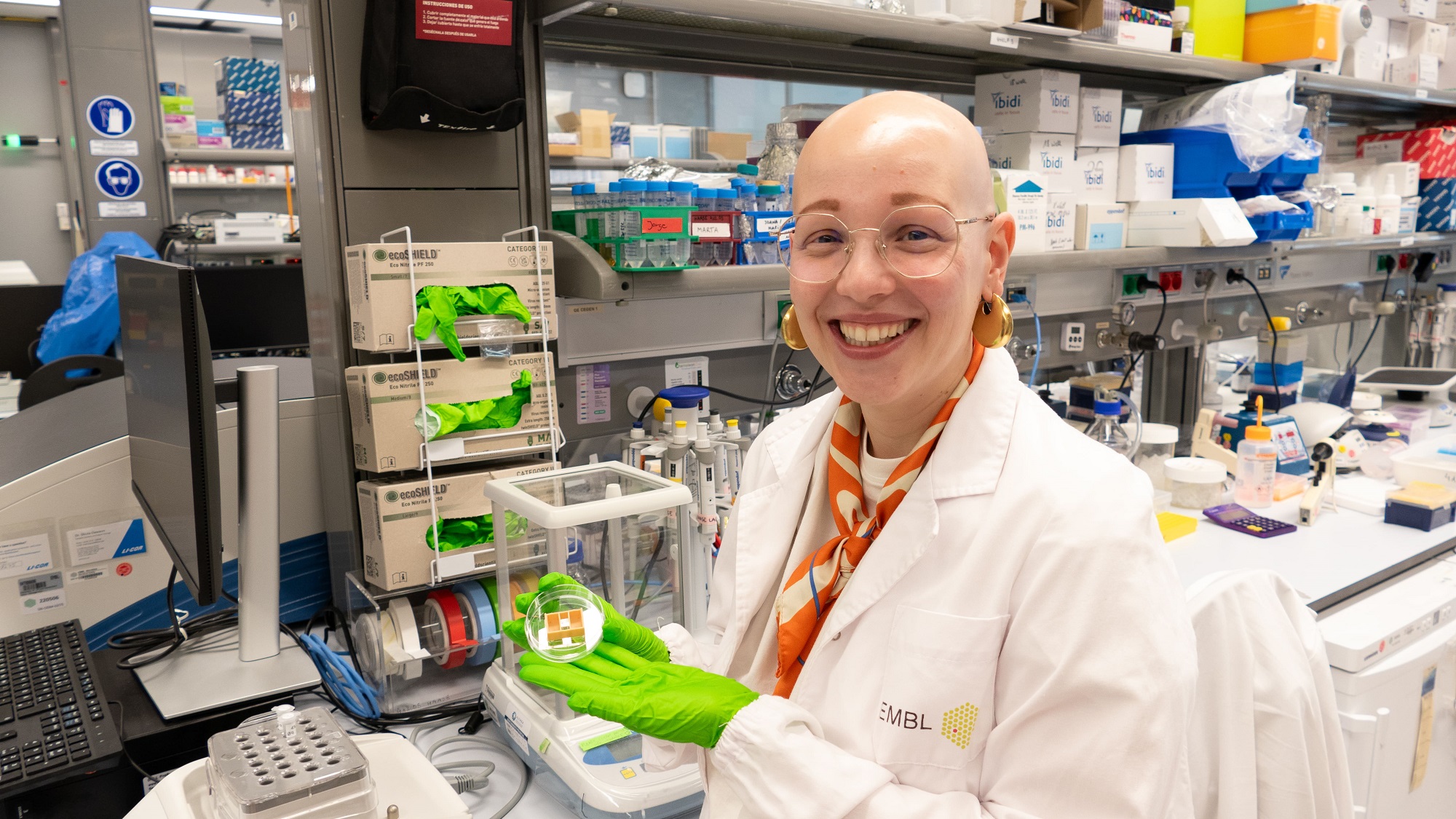
Carmen Moccia, first author of the article, tells us about the importance of using tissue-specific cells for creating the chip, as “endothelial cells have different properties and functions depending on the tissue”.
We have to use tissue specific cells, in this case from breast, to generate a mammary vascularized tissue model.
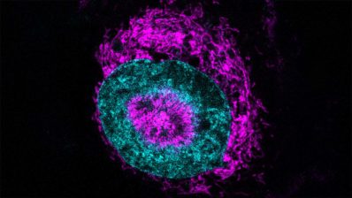
To simulate the different phases of the cycle, they added varying concentrations of estrogen and progesterone, two of the sex hormones, to the tissue on-chip. They then measured any changes in the microvessels (arterioles, metarterioles, capillaries, and venules).
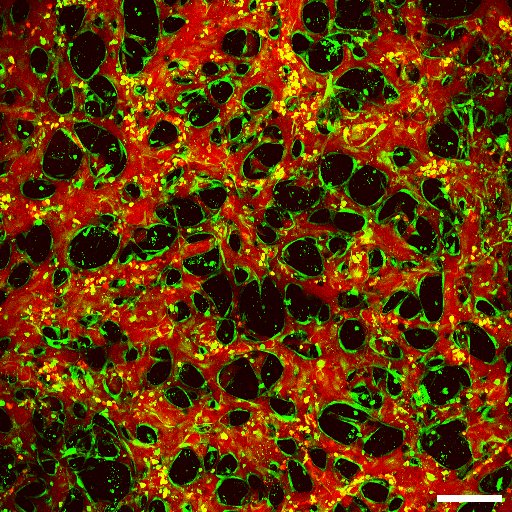
Specifically, they found that after menstruation, during the follicular phase, when hormone levels are increasing, the microvessels increase their branching and connections. Then, mimicking ovulation – with high levels of estrogen – the microvessels increase in diameter and area. Finally, mimicking the luteal phase, when progesterone levels rise, there is a progressive regression of the vessels. The barrier function of these in vitro vessels is disrupted and vessels are not perfusable at this stage of the cycle.
Our study is the first to use combined hormones in physiologic levels found in plasma to mimic levels found in the adult female menstrual cycle.
Estrogen has a multifactorial effect on the vessels: promoting the release of substances that stimulate vasodilation and vessel relaxation and reducing the production of vasoconstrictors. The role of progesterone on vascular remodeling has been under-studied, and causes extensive remodeling at high levels, as in the luteal phase. These affects have been observed in breast involution (the change from a lactating to a non-lactating breast). This research highlights the importance of using 3D models and microfluidic chips to mimic specific tissues, in this case mammary, to study the role of sex hormones in vascular remodeling.
In the future, these vascularized mammary mini-tissues could be used to study the effects of hormones on microvessels during pregnancy or following menopause. Cancerous mammary tissue on a chip is planned as a future study in the group – to assess how therapies are transported through the blood to breast cancers.
C. Moccia, M. Cherubini, M. Fortea, A. Akinbote, P. Padmanaban, V. Beltran-Sastre, K. Haase. (2023). Mammary Microvessels are Sensitive to Menstrual Cycle Sex Hormones, Advanced science, 10(35), 2302561. DOI: 10.1002/advs.202302561