This immunofluorescence image was sent to us by Pierre Bercier, Ph. D., a postdoc at the Adel Al Jord lab at the Centre for Genomic Regulation (CRG). This lab studies the mechanisms of organelle remodelling in cells – in particular, how fluctuations in physical forces, which agitate the inside of a cell, impact genetic messages and cellular function.
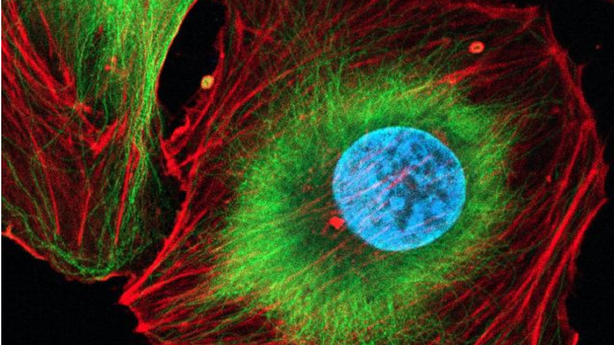
“In the image, you can see a mesenchymal cell, a type of cell found in connective tissues that can differentiate into bone, cartilage, and fat cells. The picture was taken after immunofluorescence staining, a technique that uses fluorescent dyes to label specific structures inside the cell.
The DNA in the nucleus is labeled in blue, while the cytoskeleton, the internal scaffold that gives the cell its shape and allows it to move, is shown in green and red.
More specifically, microtubules, which act like intracellular highways for transport and structural support, are labeled in green and are mostly organized around the center of the cell, forming a dense network around the nucleus. In contrast, actin filaments, which help the cell move and maintain tension, appear in red and are concentrated near the cell edges, outlining its contour.
This even distribution of cytoskeletal elements around the nucleus highlights how the cell maintains its internal organization and mechanical balance.
The image was taken using a confocal microscope, a high-resolution fluorescence microscope that captures thin optical sections of the cell, allowing 3D reconstruction, as part of a project aiming to understand how the cytoskeleton’s dynamics influence the movement and positioning of the nucleus”.