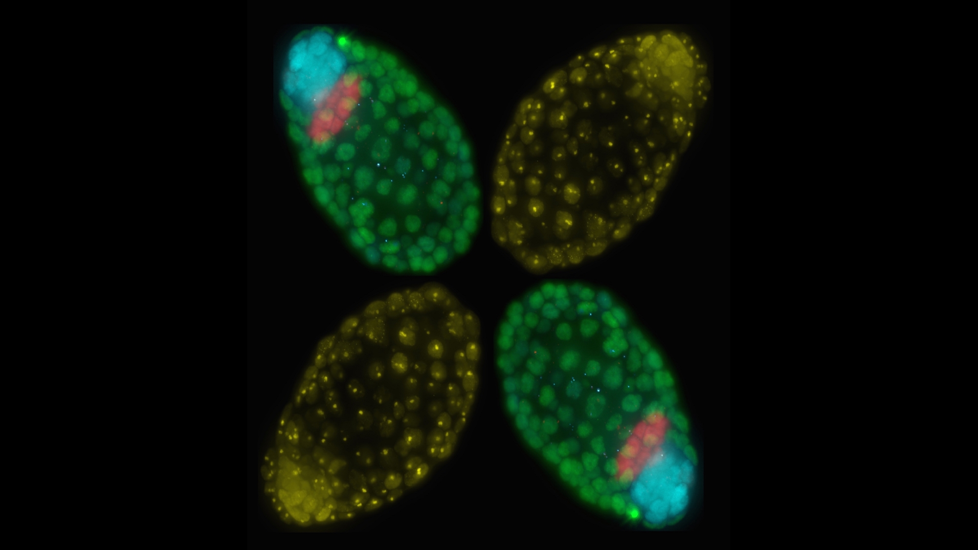
This microscope image is from Bernhard Payer’s lab at the Centre for Genomic Regulation (CRG), where they study how epigenetic information is reprogrammed during mammalian development, using the X-chromosome as a model system.
In the image, four female mouse blastocyst embryos are arranged in the shape of an X, symbolising the changes happening to the X-chromosome at this stage (4.5 days of embryonic development). Female embryos inactivate one of their two X-chromosomes (marked by the yellow foci in the picture), in order to achieve equal X-chromosome expression with males, who have only one X and one Y chromosome. In the red cells of the primitive endoderm, the future yolk sac, and the green cells of the trophectoderm, the future placenta, female mouse embryos keep the X-chromosome they inherited from their father inactive.
On the other hand, the blue labelled cells of the epiblast, which will form most tissues in the post-implantation embryo, have gone through X-chromosome reactivation, so both X-chromosomes are active. Once the embryos are implanted into the uterus, they will undergo random X-inactivation, where either their father or mother’s X will be inactivated.
The image was fittingly named “Dealing with Gender Balance” by PhD student Jacqueline Severino, and it won the second runner up prize at the Abcam Image competition 2017.
Would you like to see your photo here? Please send us pictures related to science or the PRBB to ellipse@prbb.org.

