The Al Jord lab at the Centre for Genomic Regulation (CRG) is interested in how the two major cell compartments, the cytoplasm and nucleus, communicate via physical stimuli.
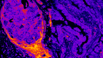
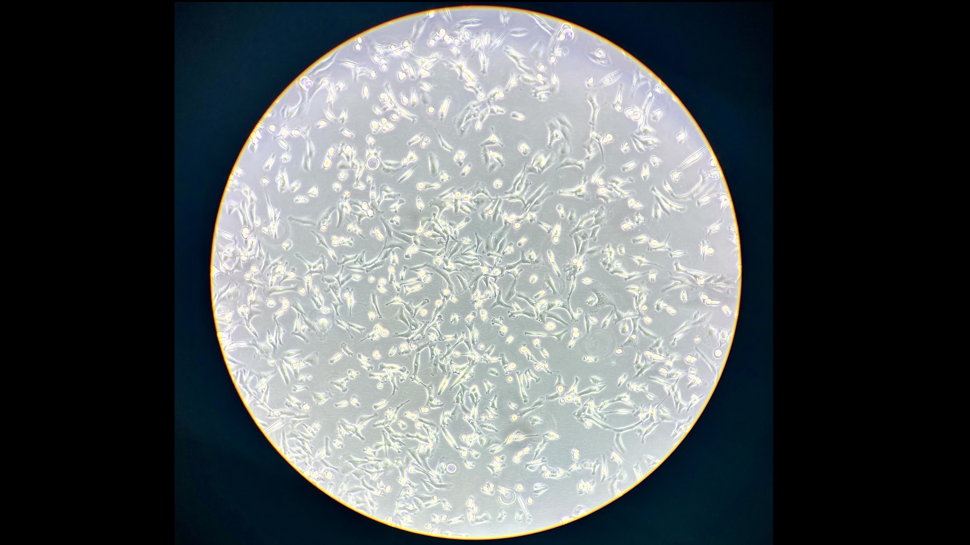
Typhaine Esteves, Lab Manager of the group, took this image with an iPhone 15 through a stereomicroscope lense. In it we can see malignant breast cancer cells prepared for a live cell experiment.
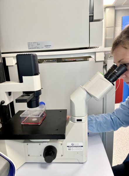
“I wanted to show what the cells look like when viewed under the microscope in routine observation. I took this photo with my phone through the microscope’s eye before preparing the cells for the experiment”, explains Thyphaine.
The experiment she was preparing aims to monitor, at high spatiotemporal resolution (imaging every 0.5 seconds for up to 60 minutes), how the cytoskeleton in the cytoplasm physically impacts molecular dynamics inside the cell nucleus.
The team will then compare the dynamics in metastatic cells to healthy cells, as well as alter the cancerous cells pharmacologically and compare them to non-altered ones.
We’re looking forward to hearing more about the results of these experiments!
Want to see your photo here? Send us images related to science or life in the PRBB to ellipse@prbb.org.