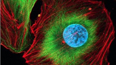
A cell is like a small city in which molecules are transported from one place to another to carry out their functions. At certain times, it is necessary to create and assemble various proteins that function as machinery capable of carrying out certain processes that are indispensable for life. For example, when DNA needs to be duplicated before cell division. This is why we cannot study cells through static images. “In the end, a cell is a bit like an engine; if you only have a picture, you don’t understand that the pistons need to move for the engine to work,” says Oriol Gallego. He is the group leader of the Live-cell Structural Biology Lab, at the Department of Medicine and Life Sciences (MELIS-UPF).
In this episode of “La mare de la ciència” Gallego explains how they are developing new microscopy techniques to study how cells work. In order to study cells in movement, they have to combine different techniques and develop new ways of looking. One example is the technique they have created to locate proteins inside the cell. Like a GPS, they use three reference points that they have introduced into the cell to triangulate the position of the molecules they are studying.
“If you want to be the first to observe a biological process and learn about biology that we don’t yet know about, you need to be very creative”
Oriol Gallego, MELIS-UPF
Listen to the podcast to discover these microscopy techniques and what different types of yeast, such as beer yeast or a Pyrenean variant, are used for in this laboratory.
We also talk about how the availability of certain tools marks progress in research; the benefits of dedicating time to university teaching; and the similarities between artists and scientists. “If you want to be the first to observe a biological process and learn about biology that we don’t know yet, you need to be very creative,” explains Gallego.
Don’t miss it!


